Latest Discovery – (2) Bilateral Coronoid Foramina: Beyond Imagination
A researcher from Bangalore, Dr. Nyer Firdoose Chintamani Subhan, has recently published his original research paper in an international journal, Journal of Surgical and Radiological Anatomy. Dr. Nyer, a maxillofacial and cleft surgeon, discovered the presence of bilateral coronoid foramina along with accessory foramina over the lateral aspect of the ramus of the mandible, Bilateral Coronoid Foramina. This unique discovery has reinforced the fact that the human body is full of surprises.
Awareness about such foramina is important as they can lead to diagnostic and therapeutic errors. This maybe one of the factors causing failure in anesthesia in dental and maxillofacial practice. Prior knowledge about presence of such anatomical variations can help avoid adverse complications while performing surgical procedures.
It was an accidental finding! Bilateral Coronoid Foramina
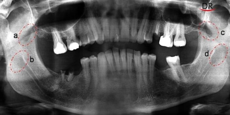
During routine examination of a patient having history of reduced mouth opening, a panoramic radiograph was taken. Panoramic radiograph revealed an unusual and peculiar finding in the coronoid process and the ramus of the mandible.

Radiographs showing a strange radiolucency on coronoid process bilaterally (a, c) as well as on ramus of mandible(b,d) which did not correlate with normal radiolucency of lingual fossa.
Author carried out further investigations by 3D CBCT, CT scan and MRI for the discovery of Bilateral Coronoid Foramina
Analysis of these scans confirmed the structural alterations like presence of large foramina/ bony defects in the bilateral coronoid processes of the jaw along with the bilateral accessory foramen on the lateral aspect of the ramus, which was not consistent with the normal morphology of the mandible. This also emphasizes the importance of complementary investigations in the diagnostic phase.


3D CBCT images confirming the presence of structural variations in the coronoid process as well as in the lateral aspect of ramus of the mandible. (a coronoid foramina; b accessory foramina on the lateral aspect of mandible; c condyle; z zygoma; mf mandibular foramen on the medical aspect of mandible; m mastoid air cells; e external auditary meatus)

CT scan images also confirming the presence of coronoid foramina and accessory foramina as above mentioned.
- Coronal section – Rm right mandibular canals; Lm left mandibular canals.
- Sagittal section left side showing: a coronoid foramen; b accessory foramen on the lateral aspect of ramus of mandible;c condyle; e external auditary meatus; m mastoid process.
- Axial section showing: Ra right coronoid foramen; La left coronoid foramina

MRI scan images showed similar structures.
- Coronal section showing neurovascular bundle of the coronoid foramina and accessory foramina on the lateral aspect of ramus. Ra right coronoid foramen; Rb right lateral accessory foramina; La left coronoid foramen; Lb left accessory foramina.
- Sagittal section left side showing: a coronoid foramen neurovascular bundle; b lateral accessory foramina neurovascular bundle.
- Axial section showing: c condyle; a coronoid foramina contents; d coronoid process foramina.
This presence of such foramina was never recorded before. These foramina are ‘nonmetric variants of the neuro-musculature’. The non-metric variants mean traits which are discontinuous or discrete in the normal anatomy of the bones.
These cannot be measured or recorded on a present or absent basis. They are genetic in most of the cases. Such abnormalities cannot be observed easily until and unless they are associated with any problems in the individual. Even in the reported case, it was an accidental discovery.
These anatomical variations might be due to embryonic developmental errors or persistence of normally obliterated structures. Still there is need of extensive research for a proper definitive conclusion as mentioned in the case report.
Ignorance is not always bliss, especially in healthcare sciences. Dr. Nyer’s discovery reinforces the famous quote –
‘what the eye doesn’t see and the mind doesn’t know, doesn’t exist’ – even if it does!
The entire team at DentalReach congratulates Dr.Nyer Firdoose Chintamani Subhan for his unique and original research work. We hope that Dr. Nyer will come up with more extensive research on the same and we wish him success on his future endeavors!
References
- Nyer Firdoose Chintamani Subhan. Bilateral ‘coronoid foramina’ with accessory foramina on the ‘lateral aspect of ramus’ of mandible: an unseen variance discovery in humans. Surgical and Radiologic Anatomy https://doi.org/10.1007/s00276-018-1984-6.18-1984-6.


Comments