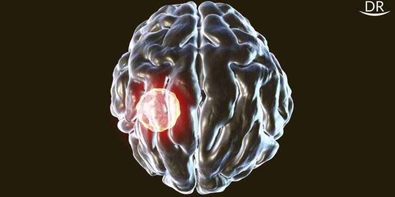
A brain abscess is defined as an abscess caused by inflammation and collection of infected material coming from local (ear infection, dental abscess, infection of paranasal sinuses, infection of mastoid air cells of the temporal bone, epidural abscess) or remote (lung, heart, kidney, etc) infectious sources within the brain tissue.
Symptoms highly suggestive of a brain abscess –
1. Fever
2. Increased intracranial pressure due to the space occupying lesion causes
- Severe consistent headache
- Vomiting
- Confusion
- Coma
3. Focal neurologic finding
- Hemiparesis
- Aphasia
- Poor co-ordination
4. Changes in mental status
- Drowsiness and lethargy
- Irritability
- Poor mental focus
- Poor responsiveness
Symptoms may worsen at night as supine position increases the intracranial pressure with complaints of a stiff neck and in occasional cases, along with blurred or double vision.
What are the causes of brain abscess?
It most commonly occurs due to a bacterial or fungal infection, though parasites might also be the cause.
When any one of these organisms enter the brain or part of the brain, it results in inflammation and swelling. An abscess will form due to the collection of infected brain cells, white blood cells and the responsible organisms.
If the abscess swells, the skull bones being non-yielding cannot expand to accumulate the growing abscess. This causes pressure on the surrounding brain tissue. This pressure can block the blood vessels preventing oxygen from reaching the brain, which results in damage to the delicate brain tissue.
How does the infection reach the brain?
There is a protective blood brain barrier guarding the brain from unwanted toxins from the blood. This barrier can sometimes be damaged due to infection causing gaps within it, thus making the brain vulnerable to pathogenic organisms.
The main routes through which an infection can enter the brain are –
- Blood through the infection from some other part of the body
- Spread from nearby site
- Result from a trauma or surgery
Infection from blood reaches the brain by crossing the blood brain barrier. In such cases, it is necessary to find the primary lesion to prevent its future recurrence.
A person with a weakened immune system is mostly susceptible to blood borne infection of the brain. The most common infections known to cause brain abscess are:
- Endocarditis
- Pnuemonia
- Bronchiectaisis
- Abdominal infections
- Cystitis
- Pelvic infections
Other than this, an infection which starts form inside the skull, like an infection of the nose or ear can also spread to the brain. Some of the examples of these type of infections include
- Otitis media
- Sinusitis
- Mastoiditis
- Untreated Dental Infection
Direct trauma is also a common cause of brain infections which can result from either a penetrating brain injury or a neurological surgery. Examples of such trauma are
- Compound skull fracture resulting in pushing of brain segments into the brain
- Presence of foreign body, such as a bullet
Diagnosing a Brain Abscess
If you see any of the symptoms mentioned above, immediately refer the patient to a neurologist who may prescribe the following tests
- MRI
- CT scan
- CT guided aspiration
- Blood tests
Possible treatment of a Brain Abscess
The first step will usually be the prescription of broad spectrum antibiotics as it may be a life threatening condition. The neurologist will alter the medications as per the responsible organisms after the confirmation from the tests. If the pressure from the abscess continues to rise and does not respond to medication, then the next viable option is surgery.
Now, why all this havoc for a dentist? How do teeth come into the picture in a life threatening condition like this?
You all must have heard about an odontogenic infection advancing to osteomyelitis or cellulitis or myofascial space infection, but a brain abscess?
Well, here are some case reports from the literature in which a simple carious lesion or post dental treatment sequelae lead to a brain abscess and in some cases proved to be fatal.
Case 1
A 53 year old female patient reported to Sun Dental Hospital, Korea with trismus and headache being her chief complaint for which she was receiving treatment from a local dental clinic since two weeks.
Clinical examination revealed facial swelling and trismus, with mouth opening < 20 mm and localised chronic periodontitis of maxillary right posterior region. Panorex showed severe alveolar resorption of 17. Laboratory tests revealed elevated white blood cells. The patient displayed elevated body temperature, myalgia, altered orientation and speech difficulty.
Imaging analysis showed a 1.5*1.5 cm perforation of right sphenoid bone and MRI showed a capsulated regular mass in the right temporal lobe with irregular edema.
The patient was diagnosed with a brain abscess of dental origin and was treated with craniotomy procedure and intravenous antibiotic therapy.
One week after craniotomy, the patient’s headache disappeared and mouth opening increased following which the upper right second molar was removed which was the suspected source of infection.
Case 2
An 11 year old boy came to a community hospital emergency department with a 2 week history of dull continuous headache and 1 week history of nausea and vomiting. He was discharged with the differential diagnosis of influenza or migraines with supportive therapy.
The following day he was found lying on the floor, screaming, holding his head, mumbling and unable to speak coherently. He also had the following symptoms

- Acute onset of confusion
- Lethargy
- Unstable gait
- Neck stiffness
MRI showed a thin walled lesion in the left temporal lobe with surrounding edema. He was immediately started on dexamethasone, mannitol, vancomycin and ceftriaxone. The boy had no history of recent sinusitis, otitis media or upper respiratory tract infection. He just reported a toothache before 3 weeks which subsided on its own. The boy underwent a craniotomy and ultra sound guided aspiration of the abscess without complication. Broad spectrum antibiotics was continued was prescribed along with phenytoin, as he reported of a ‘funny feeling’ and had a hemifacial droop on the right side which was suggestive of partial focal seizure.

Dental examination revealed grossly decayed lower left second primary molar with evidence of dental abscess which was extracted under general anesthesia.
Follow up imaging showed recurrence of the abscess on the left side which was treated by second craniotomy. Three months after his initial presentation he was discharged but continued rehabilitation under care of speech and language pathologists and occupational therapist.
Case 3
This case was diagnosed only at at post-mortem of a 30 year old male patient who died of a massive brain infection several days after the completion of root canal treatment of maxillary molar.

The patient was reportedly in good health before his general dentist performed a root canal on his maxillary left first molar without any apparent incident.
The next day patient called his dentist complaining of midfacial swelling and pain arising from the treated molar to which he prescribed amoxicillin and a moderate strength analgesic. Within 48 hours the pain and swelling had increased and the patient lost vision of his left eye. The patient was admitted to the emergency department with severe orbital proptosis and infection of the left eye. Emergeny treatment consisted of an arterial cut down and broad spectrum antibiotics.

Serial tomography suggested cavernous sinus thrombophlebitis. The patient died of this massive infection several hours after admission.
The post obturation radiograph shows a large amount of filling material from the root canal, extruded into the maxillary sinus which might have resulted in extrusion of infected material from root canal into the maxillary sinus. This resulted in maxillary sinus infection spreading to brain.
The likely pathway of spread of infection would be from maxillary sinus through the pterygoid plexus, facial vein, supra orbital vein into the cavernous sinus. The relatively direct route would account for rapid development of symptoms and relatively sudden death of the patient.
Conclusion
It is necessary to take proper post operative radiographs and prompt action with any unwanted post operative patient complaints.
Diagnosis of a dental cause of brain abscess is done by exclusion of other possible causes of infection i.e. when –
- No other source of infection is found
- Microbiological spectrum contains the oral microflora
- Clinical and radiographic signs and symptoms of an acute or chronic dental infection are present
Proper and timely treatment of odontogenic infections prevent potentially life threatening conditions. Although incidence is low, dental cause should also be considered and proper clinical examination done.
References
- Muzumdar D, Jhawar S, Goel A. Brain Abscess: An Overview. Int J Surg.2011:9(2):136-44
- Hibberd CE, Nguyen TD. Brain abscess secondary to a dental infection in a 11 year old child: case report. J Can dent assoc.2012;78:c49
- Mitchell Levine: Understanding How a Dental Infection May Spread to the Brain: Case Report J Can Dent Assoc 2013;79:d9


Very useful information and very nicely explained by case presentation.
Thank you!