– Dr. Garima Poddar
Introduction
The target of an endodontic treatment is to prevent and intercept pulpal / periradicular pathosis, to conserve a tooth affected by pathosis and to prevent recontamination after the treatment. In order to do so, a sound knowledge of basic root and root canal morphology as well as possible variation in it is important for doing a successful root canal treatment. Successful negotiation of a varied root canal anatomy followed by cleaning, shaping, and obturation of the entire canal system in 3 dimensions plays an important role in good prognosis of a tooth which has been endodontically treated.
Weine classified premolar root canal configurations into 4 types:
- Type I—single canal from pulp chamber to apex;
- Type II—two canals leaving the chamber and merging to form a single canal short of the apex;
- Type III—two separate and distinct canals from chamber to apex;
- Type IV—one canal leaving the chamber and dividing into two separate apical foramina.
Negotiating such splits and varied anatomy requires good understanding of these variations along with skill and suitable armamentarium.
This case report describes the successful diagnosis and treatment of mandibular second premolar with a Type IV Weine’s configuration.
Case Report
Clinical Presentation
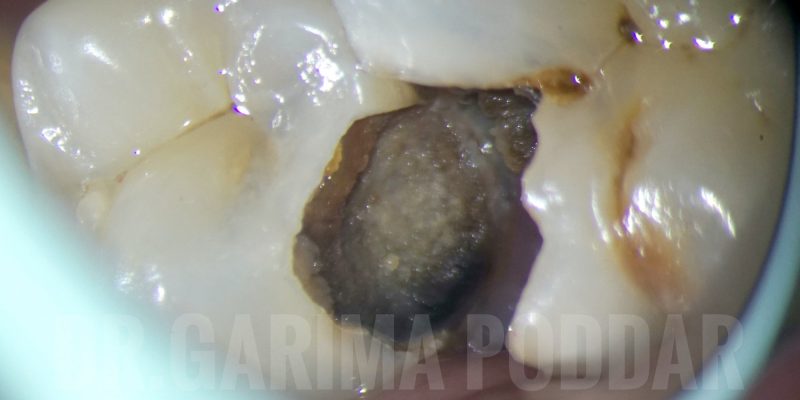
A 36-year-old female patient reported to our hospital with severe pain and tenderness in mandibular right posterior region since 15 days. On clinical examination, deep caries was observed in tooth number 45 and 46.

Radiographic examination confirmed that caries was approaching pulp in both 45 and 46. 45 showed severe tenderness to percussion.

Radiographic evaluation revealed the following-
1. Deep disto proximal decay in 45 involving pulp.
2. One canal splitting into two in the middle third region of the root in 45 – That is, having a 1-2 configuration/ type IV Weine’s configuration.
Treatment – RCT was planned for 45 first, followed by 46.
Access opening –
The tooth was isolated using rubber dam isolation and caries was excavated completely. Access prep was done and split were negotiated using 10 k hand files. Proper tactile examination and detailed study of the preoperative radiograph proved to be helpful.

Shaping protocol
- Coronal flaring was done using One flare file (Micromega, France).
- 10k file was used to instrument the canal till in became loose in both the splits.
- Glide path preparation was done with the help of One G file (Micromega, France).
- Shaping of both the canals/splits was done with 2 shape files (Micromega, France).
- The main trunk was shaped till 25-06% and rest of the canal till working length was shaped till 25-04%.
Irrigation protocol
Throughout shaping, 5.25% sodium hypochlorite was used after each file. Side vented 30 gauge needles were used for irrigation.
After shaping, the following protocol in each canal, was used for irrigation and activation of irrigants-
- 17% EDTA – 1 ml per canal – ultrasonic activation (EndoUltra, Vista).
- Distilled water used to flush the canals.
- 5.25 % sodium hypochlorite – intracanal heating of sodium hypochlorite- ultrasonic activation. (4 such cycles repeated per canal)
- Distilled water.
Obturation protocol
Canals were dried with paper points. Cone fit intra oral radiograph was made.
One split was blocked by inserting a paper point and gutta percha cone was inserted in the other split, and then the paper point in the other split was also replaced with cone. Canals were obturated with warm vertical compaction technique (EQ-V, Meta biomed). Sealer used was a resin-based sealer (Ah plus, Dentsply Maillefer).

Post endodontic restoration
Post and core build up was done for 45 using a fiber post of size 1.1 (Tenax fiber post, Coltene). The core build up was done using core build-up material (Paracore, Coltene). A full coverage crown is to be done after endodontic treatment of 46 completed.

Discussion
A sound knowledge of root canal anatomy is important for success of endodontic treatment. Also, having a fair idea about the variations in that anatomy proves to be helpful in successfully treating a tooth.
Angulated radiographs play an important role in deciphering a lot of details about the canal anatomy. A good angulated pre-operative radiograph serves to be very helpful in endodontic treatment of such teeth.
Using magnification and good source of illumination helps in treating such teeth in a conservative manner, because of better visibility.
Determining the correct working length of each canal is a very important step in a root canal treatment. Thorough cleaning, shaping and then a three dimensional obturation of canals till the exact working length, plays a vital role in a good prognosis of a tooth being endodontically treated.
Conclusion
Awareness of the variations related to configuration of canals and types in mandibular premolars is important for a clinician. Tactile examination is one of the vital steps in locating a split along with good interpretation of a pre-operative radiograph. Use of magnification is also important for such cases.
References
- F. S. Weine, Endodontic Therapy, Mosby-Yearbook, St Louis, Miss, USA, 5th edition, 1996.
- B. M. Cleghorn, W. H. Christie, and C. C. S. Dong, “The root and root canal morphology of the human mandibular second premolar: A Literature Review,” Journal of Endodontics, vol. 33, no. 9, pp. 1031–1037, 2007.
- R. R. Slowey, “Root canal anatomy. Road map to successful endodontics,” Dental Clinics of North America, vol. 23, no. 4, pp. 555–573, 1979.
- B. Dadresanfar, Z. Khalilak, and S. Shahmirzadi, “Endodontic treatment of a maxillary first premolar with type IV buccal root canal: a case report,” Iranian Endodontic Journal, vol. 4, no. 1, pp. 35–37, 2009.
Disclaimer – The views and opinions expressed in this article are that of the author alone and does not necessarily reflect the official policy of DentalReach. DentalReach does not endorse, promote or associate with this product and this article is meant for informative purposes only.


Amazing job as always..