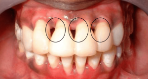
Details such as the GZP (Gingival Zenith Point), the most apical point of the free gingival margin of the periodontium, and the GZL (Gingival Zenith Length) of the lateral incisor relative to the central incisors and canine teeth can significantly influence the esthetic appearance of a smile. However, these studies, though discussing various aspects related to the gingival contours of the maxillary anterior teeth, have presented conflicting information on where the GZP should be. The appropriate placement of the gingival zenith is critical, as it helps to determine the desired axial inclination of the tooth by maneuvering the line angle of the tooth vertical axis. Subsequently, knowing the GZP of each maxillary anterior tooth from the VBM (vertical bisected midline) as well as the GZL of the lateral incisors can help facilitate a reference point during esthetic periodontal plastic surgery procedures.
The interface between tooth and soft tissues is of critical importance for achieving better esthetics.
"Inform before you perform. No surprises!"
Patient’s gingival aesthetic contribute to an improved self image and enhanced self – esteem.
Don’t change what is right to fix what is wrong. - Peter E. Dawson.
Zenith assessment should be integrated with the facial midline, smile arc, incisal plane, tooth proportions, papillary fill and periodontal phenotype. An apparent zenith discrepancy may arise from altered passive eruption, recession, tooth position, wear, restoration contour or asymmetrical gingival inflammation. The cause determines whether correction is periodontal, orthodontic, restorative or combined.
It is the most apical point of the marginal gingival scallop. Its mesiodistal position varies by tooth type and individual anatomy rather than following one universal rule.
Symmetry is desirable in a high smile line, but treatment should respect natural variation, periodontal health, tooth position and the overall composition of the smile.
It may be appropriate when excess gingival display or altered passive eruption is present, provided the bone level, supracrestal tissue attachment and final restorative margin are carefully planned.
Orthodontic intrusion, extrusion, torque and root-position changes can influence gingival margin levels and should be considered before irreversible periodontal or restorative correction.
High-quality photographs, digital scans, diagnostic wax-ups and clearly marked proposed gingival levels help communicate the treatment plan.
When tissue reconstruction is not feasible, see the role of a prosthetic gingival replacement.
Read the DentalReach dental magazine for professional perspectives, browse clinical dental articles for practical learning, and follow dental news for updates across the profession. You can also read our dental magazine editions.
Continue learning through dental events and conferences, listen to dental podcasts featuring professional conversations, and exchange questions and experiences in our dental forums.
For your next career step, explore dental jobs and opportunities across dental practices and organizations.