
GV Black Classification: Understanding the Foundations of Cavity Classification In dentistry, understanding the types of dental caries (cavities) is crucial for accurate diagnosis and treatment. One of the most widely recognized systems for categorizing dental caries is the GV Black classification. Developed by Dr. Greene Vardiman Black in the early 20th century, this classification system has been a cornerstone in dental education and practice. Dr. Black, often referred to as the "Father of Operative Dentistry," aimed to standardize the terminology and treatment methods for cavities.
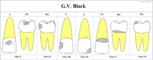
What is GV Black classification?
The GV Black classification system organizes dental caries into six distinct classes based on their location and the tooth surfaces they affect. Each class has its specific considerations for diagnosis and treatment. This system is simple yet effective in guiding dental practitioners in identifying, diagnosing, and treating various types of cavities.Class I: Cavities in pits and fissures
Class I cavities are typically found in the grooves and pits of the teeth, most commonly on the occlusal surfaces of molars and premolars. These cavities can also occur in the buccal (cheek-facing) or lingual (tongue-facing) pits of molars, as well as on the lingual surfaces of anterior teeth.Characteristics:
- Found in the natural pits and fissures of teeth
- Primarily affects the chewing surfaces of the molars and premolars
- May also be present on the lingual surfaces of incisors
Treatment:
The treatment for Class I cavities typically involves the removal of decayed tooth material and the placement of a filling. Depending on the size and depth of the cavity, restorative materials such as composite resin or amalgam may be used.Class II: Cavities on proximal surfaces of posterior teeth
Class II cavities affect the proximal (side) surfaces of posterior teeth, such as molars and premolars. These areas are located between teeth and are more difficult to clean, making them susceptible to decay.Characteristics:
- Found on the mesial or distal surfaces of posterior teeth
- Involves two or more surfaces of the tooth
- Often extends below the contact point between adjacent teeth
Treatment:
Treatment for Class II cavities requires more careful attention due to the involvement of multiple surfaces. The dentist may need to create an access point to reach the decayed area. After decay removal, the cavity is restored using a filling material, often composite or amalgam.Class III: Cavities on proximal surfaces of anterior teeth (not involving the incisal edge)
Class III cavities occur on the proximal surfaces of anterior teeth (incisors and canines) but do not extend to the incisal edge, which is the sharp cutting edge of these teeth.Characteristics:
- Located on the mesial or distal surfaces of anterior teeth
- Does not affect the biting edge of the tooth
- Can be challenging to detect without radiographs or close visual inspection
Treatment:
In treating Class III cavities, the dentist will typically remove the decay and place a composite filling material, which matches the natural color of the tooth for a more aesthetic result. The goal is to restore the function and appearance of the tooth while maintaining its structure.Class IV: Cavities on proximal surfaces of anterior teeth (involving the incisal edge)
Class IV cavities are similar to Class III but differ in that they also affect the incisal edge of anterior teeth. This class represents a more severe type of decay, as it involves a larger portion of the tooth structure.Characteristics:
- Affects both the proximal surface and the incisal edge of anterior teeth
- Often results from trauma or severe decay
- Can compromise both the aesthetics and functionality of the tooth
Treatment:
Restoring Class IV cavities can be more complex than Class III. The dentist must address both the proximal surface and the incisal edge, often using composite materials to rebuild the tooth. In some cases, more extensive restorative work, such as veneers or crowns, may be necessary if the damage is severe.Class V: Cavities on the cervical third of the facial or lingual surfaces
Class V cavities occur on the cervical third of the tooth, meaning they are located near the gum line on either the facial (cheek-side) or lingual (tongue-side) surfaces of any tooth, both anterior and posterior.Characteristics:
- Found near the gum line on the facial or lingual surfaces of teeth
- Often related to poor oral hygiene, root exposure, or wear and tear
- Can be sensitive due to proximity to the tooth’s nerve
Treatment:
The treatment for Class V cavities typically involves the removal of decayed material followed by the placement of a filling. Dentists may choose composite resin for aesthetic purposes or glass ionomer, which releases fluoride and may benefit patients with high decay risk.Class VI: Cavities on the incisal edges of anterior teeth or the cusp tips of posterior teeth
Class VI cavities are less common and occur on the incisal edges of anterior teeth or the cusp tips of posterior teeth. These areas are prone to wear and tear, especially in patients who grind their teeth (bruxism) or have a heavy bite.Characteristics:
- Located on the incisal edges of anterior teeth or cusp tips of posterior teeth
- Can result from mechanical wear, such as grinding or clenching
- May also be caused by trauma or erosion
Treatment:
Restoring Class VI cavities involves using durable materials to rebuild the worn or damaged area. Composite resins or crowns may be used to restore the tooth’s form and function, especially in cases where the incisal edge or cusp tip has experienced significant damage.GV Black classification summary table
| Class | Typical location | Clinical distinction |
|---|---|---|
| Class I | Pits and fissures of posterior teeth; selected buccal or lingual pits | Primarily involves plaque-retentive pits and fissures |
| Class II | Proximal surfaces of premolars and molars | Posterior proximal lesion, commonly below the contact area |
| Class III | Proximal surfaces of incisors and canines | Does not involve the incisal angle |
| Class IV | Proximal surfaces of anterior teeth | Includes the incisal angle or edge |
| Class V | Cervical third of facial or lingual surfaces | Describes location; the defect may be carious or non-carious |
| Class VI | Cusp tips of posterior teeth or incisal edges of anterior teeth | Often associated with wear, erosion, trauma or caries |
Why is the GV Black classification still relevant?
Despite being over a century old, the GV Black classification system remains highly relevant in modern dentistry. Its simplicity and clarity make it a valuable tool for dental professionals worldwide. The classification helps dentists communicate effectively with colleagues, plan treatments accurately, and ensure comprehensive care for patients. Moreover, it serves as a foundation for understanding dental caries and allows for the integration of newer diagnostic tools and technologies. The system’s continued use in dental education also emphasizes its importance in fostering a strong understanding of cavity types and treatments. Although advances in dental technology and materials have led to more complex treatment options, the principles of cavity classification set forth by GV Black remain applicable.Limitations of the GV Black classification
While the GV Black classification system has stood the test of time, it is not without limitations. For example, it focuses primarily on the location of cavities rather than the underlying causes or the severity of decay. Modern dentistry has evolved to take a more holistic approach to oral health, considering factors such as the patient’s overall health, diet, and risk factors for dental decay. Additionally, advancements in radiographic imaging and diagnostic technologies have improved the ability to detect caries at earlier stages, which may not always align neatly with GV Black’s categories.Free for members
Keep reading — it's free
The rest of this article is open to DentalReach members. Create a free account, or sign in, and every article on the site opens in full.

