
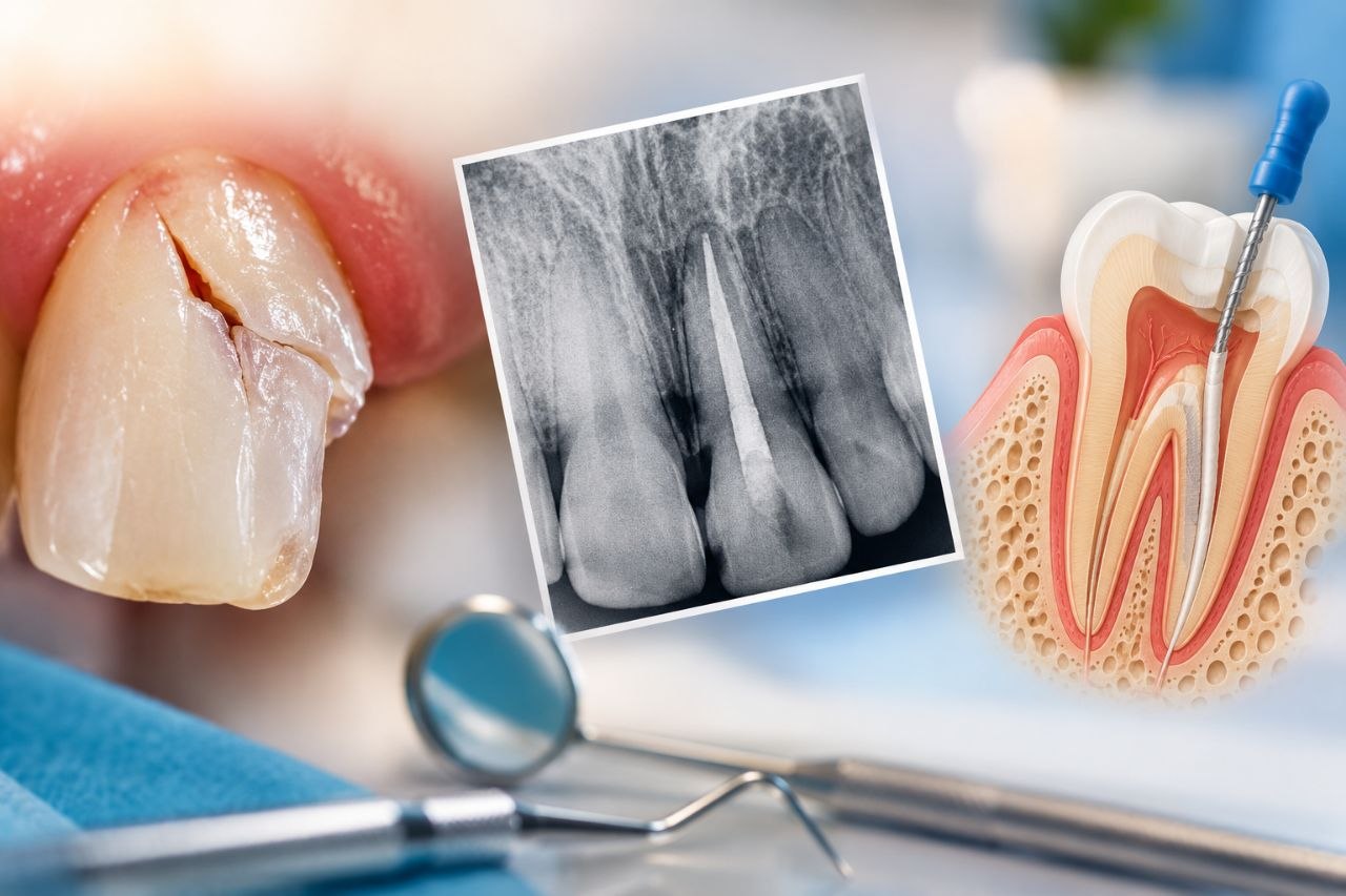
INTRODUCTION
Dental trauma is a common clinical problem, particularly among children, adolescents and young adults, who are vulnerable to falls and accidents. The maxillary anterior teeth are especially vulnerable because of their prominent position in the dental arch. Trauma can produce a wide spectrum of injuries—from minor enamel cracks to extensive crown-root fractures, root fractures, luxation injuries and complete avulsion of the tooth.
For the endodontist, the most important question is often: Does this traumatized tooth actually require root canal treatment?
The answer is not always immediate. A traumatized tooth may initially show no obvious signs of pulpal damage but may subsequently develop pulp necrosis. Conversely, a tooth with an apparently severe injury may retain pulp vitality, particularly when the root is immature and revascularization remains possible.
WHY TRAUMATIC TEETH ARE DIFFERENT
A traumatized tooth differs from a routine endodontic case in several important ways. The injury may simultaneously involve enamel, dentin, cementum, pulp, periodontal ligament, alveolar bone, neurovascular supply, root surface and surrounding soft tissues.
The prognosis therefore depends on much more than canal disinfection.
Important determinants include:
• Type of trauma
• Extent of fracture
• Pulp exposure
• Stage of root development
• Periodontal ligament injury
• Time between injury and treatment
• Extraoral dry time in avulsion
• Storage medium
• Presence of bacterial contamination
• Development of pulp necrosis
• Root resorption
• Quality of coronal restoration
• Patient compliance with follow-up
The endodontist must therefore think beyond: “Can I fill the canal?” and instead ask: “What biological events have occurred, and what can I do now to preserve this tooth?”
CLASSIFICATION OF TRAUMATIC DENTAL INJURIES
Traumatic injuries can broadly be divided into:
A. Fracture injuries: • Enamel fracture • Enamel-dentin fracture • Complicated crown fracture • Crown-root fracture • Root fracture • Alveolar fracture
B. Luxation injuries: • Concussion • Subluxation • Extrusion • Lateral luxation • Intrusion
C. Avulsion: Complete displacement of the tooth from its socket.
Each injury has a different probability of pulpal damage and therefore a different endodontic strategy.
INITIAL ASSESSMENT OF A TRAUMATIZED TOOTH
The first appointment should begin with a systematic trauma history.
History: • When did the trauma occur? • How did it occur? • Was the tooth displaced? • Was the tooth completely out of the mouth? • How long was it outside the mouth? • Was it stored in milk, saline or another medium? • Was it replanted? • Was there loss of consciousness? • Was there associated head injury? • Was there previous trauma to the same tooth?
CLINICAL EXAMINATION:Assess tooth mobility, displacement, crown fracture, crown-root fracture, soft-tissue injury, occlusion, percussion, palpation, periodontal probing, pulp sensibility, tooth colour and sinus tract.
RADIOGRAPHIC EXAMINATION: Radiographs should be carefully evaluated for root fracture, alveolar fracture, periapical pathology, root development, luxation, pulp canal obliteration and root resorption.
Additional radiographic angulations may be necessary because root fractures can easily be missed on a single projection. CBCT may be considered when conventional radiographs do not provide sufficient information and when the additional information is likely to influence treatment.
PULP TESTING AFTER TRAUMA
One of the most important clinical errors is interpreting an immediately negative sensibility test as definitive evidence of pulp necrosis.
Trauma can temporarily disrupt neural response without complete loss of pulpal blood supply. Therefore, a negative sensibility test immediately after trauma does not automatically mean that RCT is indicated.
Repeated clinical and radiographic assessment is often necessary.
Changes suggesting pulpal necrosis include:
• Persistent negative sensibility responses
• Spontaneous pain
• Presence of sinus tract
• Increasing discoloration
• Apical radiolucency
• Symptoms of apical periodontitis
The diagnosis must be based on the entire clinical picture, not a single test.
CROWN FRACTURES AND ENDODONTIC MANAGEMENT
Uncomplicated Crown Fracture:
An uncomplicated crown fracture involves enamel and dentin without pulp exposure.
Management may include:
• Reattachment of the fragment
• Composite restoration
• Dentin sealing
• Regular pulp monitoring
RCT is not routinely indicated simply because dentin has been exposed. The pulp should be monitored because traumatic pulpal damage may become apparent later.
Complicated Crown Fracture
A complicated crown fracture involves exposure of the dental pulp.
Treatment depends on:
• Patient age
• Time since injury
• Size of exposure
• Degree of contamination
• Root maturity
• Pulpal status
Vital pulp therapy may be preferable to conventional RCT in appropriate cases. Possible procedures include direct pulp capping, partial pulpotomy and full pulpotomy.
This is particularly important in immature permanent teeth because preservation of vital pulp tissue can allow continued root development.
Therefore: Pulp exposure does not automatically equal root canal treatment.
Crown-root fractures
Crown-root fractures extend through enamel and dentin into the cementum and may involve the pulp. They are clinically challenging because the fracture frequently extends subgingivally. Treatment options depend upon fracture location, restorability, periodontal involvement and root development.
Possible approaches include:
• Fragment removal and restoration
• Endodontic treatment
• Surgical crown lengthening
• Orthodontic extrusion
• Intentional replantation in selected cases
• Extraction when the tooth is non-restorable
The endodontic treatment plan should always be developed alongside the restorative plan.
ROOT FRACTURES
Root fractures are among the most interesting trauma cases from an endodontic perspective. They may occur in the cervical, middle or apical third and may be horizontal or oblique. Importantly, a root fracture does not automatically mean extraction or RCT. Healing can occur between the fractured segments, particularly when the coronal segment is repositioned and stabilized appropriately. The pulp may remain vital. Therefore, the initial objective is often to preserve the pulp and stabilize the tooth rather than immediately perform RCT.
WHEN IS RCT INDICATED IN ROOT FRACTURE?
Endodontic treatment is generally directed toward the coronal segment when pulp necrosis develops. The apical fragment may remain vital and should not automatically be instrumented.
The clinician should determine:• Which segment is necrotic? • Is there communication between the fracture line and oral environment? • Is there infection? • Is there apical pathology? • Is the fracture located coronally? • Is the coronal segment restorable?
In selected cases, successful endodontic treatment of the coronal fragment can allow the tooth to remain functional.
AVULSION – THE TRUE DENTAL EMERGENCY
Avulsion is complete displacement of a tooth from its socket.
Unlike most dental trauma, avulsion requires immediate action because the periodontal ligament cells on the root surface are rapidly compromised outside the socket.
The prognosis depends significantly on: • Extraoral time • Extraoral dry time • Storage medium • Condition of periodontal ligament cells • Root maturity • Speed of replantation • Subsequent infection and resorption
The fundamental principle is: REPLANT AS SOON AS POSSIBLE.
WHAT SHOULD BE DONE WITH AN AVULSED TOOTH?
If immediate replantation is possible, the tooth should be handled by the crown rather than the root. The root should not be aggressively scrubbed because this may damage remaining periodontal ligament cells.
If immediate replantation is not possible, the tooth should be placed in an appropriate storage medium such as milk, physiological saline or a specialized tooth-preservation solution.
The aim is to minimize dehydration of periodontal ligament cells.
Free for members
Keep reading — it's free
The rest of this article is open to DentalReach members. Create a free account, or sign in, and every article on the site opens in full.