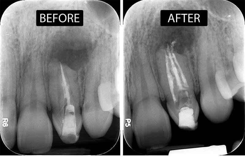
Persistent cold sensitivity in a root canal–treated tooth is uncommon, but when it does present, it can create significant diagnostic confusion. Conventional teaching suggests that once the pulp is removed, thermal sensitivity—especially to cold—should no longer be present. Yet clinically, patients occasionally report exactly that.
A recent narrative review examines this paradox and highlights the mechanisms and clinical implications behind cold-only sensitivity following root canal therapy.
From a biological standpoint, the absence of pulp does not mean the absence of sensory response. The periodontal ligament (PDL) and periapical tissues remain richly innervated. These structures contain mechanoreceptors and nociceptors capable of responding to external stimuli, including temperature changes.
In addition, neural adaptation following endodontic treatment appears to play a role. Studies have shown that after pulpectomy, neural elements in the apical and periodontal regions may undergo sprouting and reorganization, potentially increasing sensitivity to certain stimuli, particularly cold.
Another important contributor is residual or accessory anatomy.
Even in well-executed root canal treatment, small amounts of tissue may remain in lateral or accessory canals. While these remnants may not sustain vascular supply, they can still retain sensory capacity. Under certain conditions—particularly when exposed via cracks or leakage—these structures may respond to cold stimuli.
However, the most clinically relevant factor emphasized in the review is structural compromise, particularly root cracks.
Cracks alter the way thermal stimuli travel through the tooth. They can act as conduits, allowing rapid transmission of cold to deeper structures, including dentin tubules and the PDL. This results in fluid shifts and activation of mechanosensitive nerve fibers, similar to the hydrodynamic mechanism observed in vital teeth.
In such cases, cold sensitivity may be the only presenting symptom.
Radiographically, these teeth may show features such as:
Localized “J-shaped” lesions
Narrow, isolated periodontal defects
Subtle periapical changes
CBCT imaging and careful clinical examination are often required for confirmation.
Restorative factors also play a role.
Loss of coronal seal, marginal leakage, or exposed dentin surfaces can permit thermal conduction toward deeper tissues. Inflammation in the periapical region, whether due to microbial leakage or overextended filling materials, may further lower the activation threshold of sensory fibers, making them more responsive to cold.
Clinically, the challenge lies in accurate diagnosis.
Cold sensitivity in a previously treated tooth should prompt a systematic evaluation rather than immediate retreatment. Key steps include:
Isolating the tooth to rule out referred sensitivity
Assessing restoration integrity and coronal seal
Evaluating for cracks using transillumination, magnification, or CBCT
Checking for periapical pathology
Performing bite tests to identify fracture-related pain
Importantly, adjacent vital teeth must always be excluded as the source of symptoms.
Management depends entirely on the underlying cause.
Free for members
Keep reading — it's free
The rest of this article is open to DentalReach members. Create a free account, or sign in, and every article on the site opens in full.