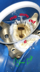
Let’s understand the basics of calcified canals!
A calcified canal is a root canal system complication, occurring due to hard tissue deposition which makes the canal narrower. Calcification is uncontrolled due to enzyme pyrophosphatase failure that results in reduction in capillary permeability and blood supply, causing calcifications. Canal calcification is usually termed as an obliteration of the pulp canal. It causes the darker hue, translucency loss and the yellowish appearance of the tooth’s crown.
What Causes a Calcified Root Canal?
AGE: In geriatric patients the incidence of calcified canals is high due to secondary or tertiary dentin formation.
LONGSTANDING TOOTH DECAY OR OLD RESTORATIONS & CROWNS: If a patient has a large amount of tooth decay, our body can shrink the pulp for protection. It means the pulp receives less blood supply. Reduced blood supply increases the risk of infection, which will require a root canal treatment.
TRAUMA: As sports activity increases among school-aged children, this has led more young patients to experience tooth trauma due to a facial injury. When a tooth suffers a blow, this may cause bleeding in the canals, which can become a focal point for calcification.
Root canal treatments in calcified canals- HOW?
Root canal treatments in calcified canals are always a challenge for operating dentist because it can be difficult to get even the smallest of tools down there.
Cleaning out a calcified root canal takes much more skill, time and patience than a standard root canal procedure.
Fortunately advances in knowledge and armamentarium helps in the management of calcified root canals, thus allowing resolution and restoration of teeth that perhaps would previously have been extracted, despite potentially being functional and restoratively sound
Steps to follow:
- Assessment - clinical and radiographic
- Magnification and Access Preparation
- Instrumentation
- Chemo-mechanical disinfection.
1. Clinical & radiographic assessment-
Teeth undergoing pulpal obliteration are generally asymptomatic. The affected teeth do not always react to sensitivity tests for some time.
Pre-operative radiograph assessment is important in such cases. For multirooted teeth it is recommended to have more than one pre-op radiograph in different angulations to help gauge the depth from the crown to the pulp chamber.
To simplify this process it is better to take CBCT of the tooth which will give 3 dimensional view
2. Magnification and Access
Magnification in the form of dental loupes or an operating microscope, is of paramount importance in terms of managing these cases. These aid in providing visualisation and illumination to highlight landmarks in the pulp chamber that can aid in location and management of calcified canals.
For access preperation try to locate white spots with the help of Endodontic DG16 (Dentsply Sirona) probe or micro-orifice openers along with ultrasonics in such canals.
In multi-rooted teeth, use of dyes can also be beneficial in terms of locating canal orifices. An example of this is Cerkameds ‘Canal Detector’, which works by having a dye contained in the product which invades into the root canal orifices and dyes them blue, thus enabling easier detection.
3. Instrumentation
Once any calcified canals are identified, negotiation of them presents another challenge.
Creating coronal flare with an orifice shaper is a great way to gain further access into a calcified system.
The small hand files are important in creating a ‘glide path.’Usually, if you use pre curved small hand files such as a size 6, 8 or 10 K-file you will then be able to negotiate further into the system
Nickel-titanium rotary instruments can be safely taken into the portion of a calcified canal where a glide path has been created with a size 15 or 20 K-file.
Alternating between rotary and hand instrumentation, while constantly bathing the pulp chamber of the tooth, can expedite negotiation to the apex.
In canals that are extremely calcified or curved, traditional stainless steel K files can struggle to negotiate the canal system and often unwind, twist and thus run the risk of fracturing. Dentsply’s (Dentsply Sirona Endodontics) C-Pilot files and MANI D-Finders are often a good alternative in such cases. These files are made from a special steel alloy with a uniform structure, which offers maximum resistance to fracture, but with no limitations with flexibility. The files have an inactive tip, which allows the instrument to follow the canal rather than cutting its own pathway, thus decreasing the risk of perforation of the root canal system.
Free for members
Keep reading — it's free
The rest of this article is open to DentalReach members. Create a free account, or sign in, and every article on the site opens in full.