
VistaScan
The conventional x-ray development and it's introduction in the field of dentistry has been the cutting edge of surgical diagnostics. Digital x-ray offer images with a high-resolution factor in order to meet the diagnostic needs for the dentists. The development of technology results by leading time from over 50 years of experience towards innovative and practice oriented solutions.
The VistaScan image plate scanner helps dentists diagnose faster. It is a compact device which is considered as easy to use, having minimum space requirement, and can be installed in the treatment room.
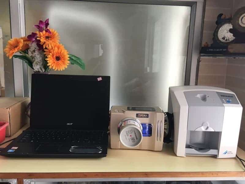
How to use it VistaScan?
The Vistascan mini model consists of sensors which are present in 2 sizes i.e 0 and 2 which is used with vista scan mini easy, while the rest come in sizes 0-4. Each comprises of 2 parts. The sensors are very thin in nature as compared to the RVG sensor. The adhesive of the sensor pack is removed and placed intraorally. The X-ray tube is positioned for shooting of X-ray. The sensor consists of 2 sides i.e printed version (written as back side above it) and plain side (X-ray tube facing side).

It has a dot which is helpful for identifying right and left sides and the midline while making X-rays.
VistaScan Scanner:
It consists of 2 lids which are of the size for sensors 0 and 2 separately, based on the sensor used for the patients.The lids have to be changed. The scanner power must be switched on by pressing the button for few seconds and it gets ready by a glowing red light .The software which is installed in the laptop/computer system must be opened and the data details have to be entered and at the end, the data must be saved.
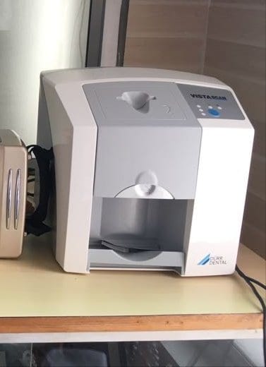
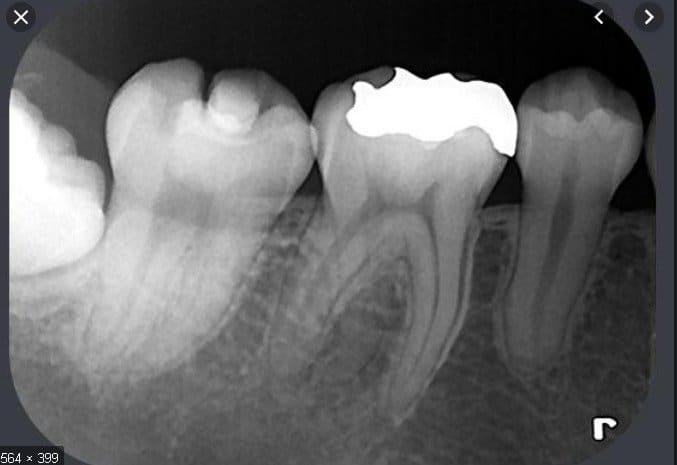
The next aspect to be followed is to click on the icon 'X-ray', and the tooth number which has to be scanned must be selected, along with the resolution data(ideally 1000 dpi sounds good). The sensor is then placed in the scanner and the scanning takes place. The X-ray image is seen on the computer screen and this image data is immediately must saved. Finally, the X-ray image can be viewed by clicking on viewer icon, providing data that is helpful for diagnostic purposes.

Free for members
Keep reading — it's free
The rest of this article is open to DentalReach members. Create a free account, or sign in, and every article on the site opens in full.