Abstract
Caries detecting dyes have been developed as clinical aids to enhance the removal of infected dentin during caries excavation, allowing for more conservative and precise operative procedures. These dyes selectively stain the denatured collagen of carious dentin without affecting healthy tissue, thereby helping in the prevention of over-excavation and potential pulp exposure. This review explores the composition, mechanism, application, indications, contraindications, advantages, disadvantages, and commercially available brands of caries detecting dyes.
Keywords: Caries detection dye, infected dentin, conservative dentistry, minimally invasive dentistry, dye-assisted excavation.
Introduction
Dental caries remains one of the most prevalent chronic diseases globally. Modern caries management emphasizes minimally invasive techniques that preserve as much sound tooth structure as possible, while ensuring the complete removal of infected tissues. Conventional caries excavation, relying solely on tactile and visual cues, can be subjective and may result in either under- or over-excavation.
Caries detecting dyes were introduced to assist clinicians in identifying infected dentin. These dyes stain the collagen matrix of irreversibly denatured dentin, facilitating its selective removal. The use of such dyes aligns with the philosophy of minimally invasive dentistry, offering a valuable adjunct in both restorative and pediatric procedures.
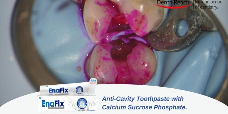
Caries detector clearly differentiates clinically the two layers of carious dentin by staining the outer carious dentin in a scarlet red color, yet not staining either the inner affected or normal dentin. The distinct staining of only the outer, infected carious layer provides a visible, clinically precise guide for caries removal.
Uses of Caries Detecting Dye
- To differentiate between infected and affected dentin.
- As an aid in minimally invasive caries removal techniques.
- In cases of deep caries where pulp preservation is critical.
- For training and education in operative dentistry.
- In pediatric dentistry to avoid pulp exposure in young permanent or primary teeth.
- To reduce reliance on subjective criteria for caries excavation.
How to Use the Dye
- After removal of superficial debris and plaque, isolate the cavity.
- Apply the dye using a micro-brush or cotton pellet to the prepared cavity surface.
- Leave the dye in contact with dentin for 10–15 seconds (varies by brand).
- Rinse thoroughly with water or air-water spray.
- Evaluate stained areas – stained dentin is considered infected and should be removed.
- Repeat the process if necessary until no stained dentin remains.
Indications
- Deep carious lesions approaching the pulp.
- Restorations in children and anxious patients where speed and safety are priorities.
- Use in conjunction with hand excavation or chemomechanical techniques like ART.
- Training purposes in dental education.
- Conservative caries removal in medically compromised or geriatric patients.
Contraindications
- Allergy or hypersensitivity to dye components.
- Shallow lesions where excessive removal is not a concern.
- Situations where cost constraints limit additional material use.
- In cases with poor visibility where staining may confuse margins.
Advantages
- Enhances precision in caries excavation.
- Prevents over-removal of healthy dentin.
- Aids in preservation of tooth vitality.
- Useful in teaching caries detection and removal.
- Non-invasive and safe when used properly.
Disadvantages
- May stain healthy dentin if not used correctly.
- Possible false positives due to non-specific protein staining.
- Does not differentiate between active and arrested caries.
- Adds time and cost to procedures.
- Risk of leaving residual dye if not rinsed thoroughly.
Common Brands of Caries Detecting Dyes in Dentistry
| Brand Name | Manufacturer | Composition | Key Features |
| Seek | Ultradent | Propylene glycol base with red dye | High visibility; easy application and rinse |
| Sable Seek | Ultradent | Propylene glycol with green dye | Green color contrasts well with red tissues |
| Caries Detector | Kuraray | 1% Acid Red 52 in propylene glycol | Reliable; widely used; consistent staining |
| Reveal | Centrix | Acid red-based dye | Syringe delivery for controlled application |
| Tooth Colored Caries Detector | GC | Transparent dye that changes on contact | Aesthetic; especially useful in anterior teeth |
| Blue Caries Indicator | Voco | Blue dye in aqueous base | High contrast for better visualization in deep lesions |
| Caries Finder | Dental Avenue | Propylene glycol base | Economical; suitable for routine use |
Conclusion
Caries detecting dyes offer a valuable adjunct in modern restorative dentistry by allowing for accurate and conservative removal of infected dentin. Their utility in minimizing the risk of pulp exposure and preserving healthy tooth structure is well recognized, particularly in deep lesions and pediatric dentistry. However, they must be used judiciously and interpreted carefully to avoid over-reliance or misdiagnosis.
Understanding their mechanism, proper usage, and limitations ensures optimal outcomes and aligns with the principles of minimally invasive dentistry. Further research into more specific and less staining formulations could enhance their clinical acceptance.
References
- Kidd EA. How ‘clean’ must a cavity be before restoration? Caries Res. 2004;38(3):305–13.
- Yip HK, Smales RJ. Caries detection techniques. Int Dent J. 1995;45(3):173–82.
- Nakajima M, et al. Relationship between caries dye-stained dentin and bacterial infection. Dent Mater J. 1995;14(1):26–33.
- Banerjee A, et al. Clinical management of carious lesions using minimally invasive techniques. Br Dent J. 2000;188(11):603–9.
- Fukuda M, et al. Evaluation of the use of a caries detector dye in caries diagnosis. Oper Dent. 2006;31(5):604–9.
- Hosoya Y, et al. Evaluation of a caries detector dye: an in vitro study. J Dent. 2001;29(7):451–6.
- Neves Ade A, et al. Effect of caries detector dyes on the microtensile bond strength to caries-affected dentin. Oper Dent. 2011;36(3):239–47.
- Russo J, et al. Evaluation of Caries Detector Dyes in Minimally Invasive Dentistry. J Clin Pediatr Dent. 2010;35(1):43–7.
- Sato Y, Fusayama T. Clinical application of a caries-detecting dye. Oper Dent. 1976;1:118–25.
- Fusayama T. Two layers of carious dentin; diagnosis and treatment. Oper Dent. 1979;4(2):63–70.
- Thomas M, et al. In vivo evaluation of the effectiveness of caries detector dye in dental caries excavation. J Indian Soc Pedod Prev Dent. 2007;25(3):134–6.
- Sridhar S, et al. Comparative evaluation of caries removal efficacy of different caries detector dyes. Contemp Clin Dent. 2011;2(3):170–3.
- McComb D, et al. In vivo comparison of caries detector dyes and removal techniques. J Am Dent Assoc. 1996;127(6):743–8.
- Massara ML, Alves JB, Brandão PR. Micromorphology of the dentin substrate in primary teeth after caries removal with a chemo-mechanical method. Caries Res. 2002;36(3):206–11.
- Ferreira Zandona A, Zero DT. Diagnostic tools for early caries detection. J Am Dent Assoc. 2006;137(12):1675–84.


Comments