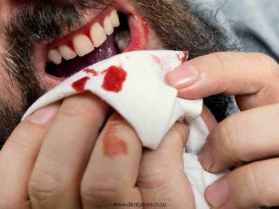
There are different types of injuries which may vary from minor damage of teeth to grossly comminuted fractures of the skull. Teeth and facial injuries are common among them. National Trauma Databank of American College of Surgeons reported 7,22,824 incidents of trauma in 2010. Falls contributed to major mechanism of injury (38.4%), followed by motor vehicle collisions (28.9%). Injuries from mechanisms of being struck (7.5%), pierced (4.7%) or related to firearms (4.5%) were less common. Head and face injuries accounted for 59.7%. Hence, head and neck trauma are major injuries in the United States.
National Crime Records Bureau (NCRB) is the principal nodal agency, that is solely responsible for the collection, compilation, analysis and dissemination of injury-related information. NCRB had reported 27,10,019 accidental deaths; 1,08,506 suicidal deaths and 44,394 violence-related deaths in 2001. Major cause of trauma was suicide (27%) followed by road accidents (20%), violence (11%), burns (9%), poisoning (6%), drowning (6%), domestic violence (2%) and falls (2%). According to the study of Hsiao et al., head and neck injuries had contributed to 60% of the road traffic injury (RTI) death list in India. In 2012, a forensic study was conducted by Pate et al to analyze the incidence, pattern, mechanism and mode of head injury among 21-30 years. Major accidents were road accidents followed by fall from height and railway. The maximum victims of head injuries were males.
Dento alveolar injuries are limited to teeth and supporting structures of the alveolus. 7-15 years of boys are mostly affected as compared to girls. Maxillary central incisor (80%) is the most commonly affected tooth followed by maxillary lateral incisor, mandibular central incisor and mandibular lateral incisor. The etiology includes road accidents, fall during infancy, sports injuries, domestic violence, iatrogenic causes to permanent tooth bud and inferior alveolar nerve, mental retardation & epileptic seizures, alcohol & drug-related injuries. Direct trauma occurs due to force applied directly to teeth and indirect trauma occurs when indirect force is applied to teeth causing the jaws to strike each other.
A dental injury represents acute transmission of energy to the teeth and supporting structures, which results in fracture and displacement of teeth and separation or crushing injuries. Dental trauma leads to damage to tooth structure, surrounding periodontal ligament, vascular and nerve supply, surrounding bone. This damage is related to the extent of displacement from original anatomic positions.
CLASSIFICATION
There are different types of dental injuries based on the classification suggested by Ellis and Davey in 1970:-
- Class I – Fracture involving enamel
- Class II – Fracture involving enamel & dentin
- Class III – Fracture involving enamel, dentin & pulp
- Class IV – Teeth that lost their vitality with or without loss of crown
- Class V – Traumatically avulsed tooth
- Class VI – Fracture of root with or without crown fracture
- Class VII – Displacement of tooth without crown/root fracture
- Class VIII – Cervical crown fracture
- Class IX – Fracture of deciduous teeth
According to Andreasen, dental injuries can be divided into:
- Fractures of the teeth
- Coronal Fractures
- Involving only enamel
- Involving enamel and dentine
- Involving enamel, dentine and pulp
- Involving enamel, dentine and cementum
- Involving enamel, dentine, cementum and pulp
- Root Fracture
- Without coronal fracture
- With coronal fracture
- Coronal Fractures
- Luxation injuries to the teeth
- Concussion
- Subluxation
- Intrusive luxation
- Extrusive luxation
- Lateral luxation
- Avulsion
- Fractures of the alveolar bone
- Fractures of socket
- Fractures of alveolar process
- Fracture of associated jaw
- Other injuries
- Displacement of a tooth which may become dilacerated
- Soft tissue injuries such as:
- Laceration
- Imbedding of a foreign body
- Latrogenic injuries such as:
- Injuries sustained during extractions, including damage to adjacent teeth and fracture of alveolar bone
- Perforation of tooth apex or side of the root during conservative or endodontic treatment
- Swallowing or inhaling of an avulsed tooth
TYPES OF TOOTH FRACTURES
Dental crown fractures contribute to 25% permanent teeth and 40% deciduous teeth injuries. Dental root fractures comprise 7% permanent teeth injuries and 3% to deciduous teeth injuries. Vertical root fractures are mostly found in 5% of total crown-root fractures.
DENTAL CROWN FRACTURES
-
-
- Most commonly involve anterior teeth
-
Fractures involving crown fall into three categories:
-
-
- Enamel without loss of substance, infraction of crown or cracks
- Enamel and dentin with loss of tooth substance but without pulpal involvement.
- Enamel, dentin and pulp with loss of tooth substance and exposure of pulp.
-
INFRACTION
-
-
- Defined as microcrack in the thickness of the enamel
- Quite common but not readily detectable
- Illuminating crowns with indirect light causes cracks
-
Management
-
-
- Routine vitality testing
- Sealing of cracks with adhesive systems
-
UNCOMPLICATED
-
-
- More common than complicated
- Usually occur at mesioincisal or distoincisal corners of maxillary central incisors
- Exposed dentin sensitivity to thermal, chemical and mechanical stimuli
- Deep, pink blush of pulp may be appreciated through the thin remaining dentin wall
-
Management
-
-
- Composite build up
- Coronal fragment reattachment
-
COMPLICATED
-
-
- Bleeding from exposed pulp
- Exposed pulp is sensitive to stimulation
- Pulp testing is positive unless there is concomitant luxation injury
- Depending on the absence or presence of a concomitant luxation injury the pulp will present with bright red, cyanotic or ischemic appearance
-
Management
-
-
- Vital pulp therapy
- Direct pulp capping
- Partial pulpotomy (Cvek Technique)
- Cervical pulpotomy
- Pulpectomy
- Follow up
-
DENTAL ROOT FRACTURES
-
-
- Maxillary central incisors most commonly affected
- Coronal fragments are displaced lingually and slightly extruded
- Degree of mobility of crown i.e. fracture plane to the apex, more stable is the tooth
- Root may occur in association with alveolar process fractures
- Temporary loss of sensitivity is seen which returns back to normal within 6 months
-
Management
-
-
- More apical and better than prognosis
- Middle and apical, reduction to position and immobilization
- Endodontic therapy when evidence of pulpal necrosis
- Coronal 3rd , Extraction
-
VERTICAL ROOT FRACTURES
-
-
- Fracture line is parallel to the long axis of the tooth and can extend from crown to the apex
- May or may not involve the pulp chamber
- Seen most often in mandibular molars
-
Clinical features
-
-
- Low level dull pain-cracked tooth syndrome
- Bite test: Pain on release of biting force
- Transillumination
- Staining with disclosing dye
- Definitive diagnosis is made only on surgical exploration
-
Management
-
-
- Single-rooted teeth: extraction
- Multirooted teeth: Hemisection with endodontic therapy and crown
-
COMBINATION CROWN AND ROOT
-
-
- Involve both crown and root
- Usually involves pulp
- Permanent teeth are more affected than deciduous teeth
- Seen often in anterior teeth due to direct trauma
-
Clinical features
-
-
- Anterior tooth: plane extends obliquely from the mid portion of the crown facially and extends below the gingival level palatable.
- Displacement of segments are minimal
- Bleeding from pulp
- Pain on mastication
-
Management
-
-
- Removal of coronal fragment permits evaluation of extent
- If pulp is not exposed: conservative treatment (Bonding tooth fragment or composite build up)
- If small amount of root is lost with pulp exposure: crown lengthening + Endodontic therapy
- If > 3-4 mm of clinical root is lost: removal of residual root
-
Bonus: Download our monthly e-bulletin!Click here to get it
DISCLAIMER : “Views expressed above are the author’s own.”


Comments