– Dr. Sejal Shah
Abstract
In a society that values appearance, those who exhibit malformed parts of the face, neck and oral cavity may become less socially acceptable. Rehabilitation of the maxillofacial patient into society requires a broad knowledge of prosthodontics, plus the capacity for compassionate patient management. Oral cancer is now a common occurrence requiring partial or complete maxillectomy. The most common oral complication of the current pandemic Covid is mucormycosis, and treatment requires partial resection of the maxilla. Rehabilitation starts with a good impression of the maxillary defect. Maxillofacial defects are always unique, and require special impression techniques, one of which is explained in this case report.
Key words-Obturator, Impression
Introduction
The cleft palate is an opening in the hard and/or soft palate. It may be genetic due to improper union of the maxillary process and the median nasal process during the second month of intrauterine development. It may also be acquired and the most common causes for maxillary defects are trauma, disease, pathological changes, genetics, surgical intervention, syndrome associated, drugs, radiation burns etc.
Primary Objectives
In the total rehabilitation of the maxillectomy patient, there are two primary objectives:
- To restore the functions of mastication, deglutition and speech.
- To achieve normal oro – facial appearance.
Impression Technique
- The small defects should be blocked out with moist cotton or gauze (gauze or cotton should be lubricated with vaseline or petrolatum).
- Larger defects with gross undercuts should be packed with 4 x 4 inch gauze squares, it should be readily retrieved and they should be shoved into the defect.
- With time, the dentist will acquire clinical judgment with regard to which areas need blocking out prior to impressions.
Step by Step Procedure
- Preliminary impressions
- Primary cast
- Fabrication of special tray
- Final impression
- Obtaining the master cast
Preliminary Impressions
The intra – oral defect can be seen in Fig. 1.

Material of choice for preliminary or primary impression – irreversible hydrocolloid impression material i.e alginate or impression compound (Fig. 2).

Diagnostic Casts
Diagnostic cast was made with plaster of Paris in a regular manner (Fig. 3).

Fabrication of Special Tray
Any undercuts in the primary cast should be blocked by using utility wax. Wax spacer adapted on the cast including the defect area (Fig. 4). Tissue stops were included to help proper application of forces. Special try was fabricated (Fig. 5).


Border Molding
Border molding the posterior and lateral area of a maxillectomy requires that the patient go through certain head and mandibular movements.
Movements to be Performed
The patient has to open and close the mouth, move the mandible from side to side, turn the head from side to side, place the chin down to the chest, move the head from side to side, and extends the head backward after seating the tray.
Border Molding
Special tray was checked in patient’s mouth and the space between the tray and tissues was examined. Tray adhesive was applied to increase the adhesion between the tray and additional silicone and border molding was performed (Fig. 6).

Final Impressions
Spacer was removed and wash impression was performed except in defect area. Putty material was mixed and secured with gauge piece and placed in patient defect area and all head moment are recorded. After recording the defect area putty was attached to special tray (Fig. 7).

Master Casts
Master cast was poured using dental stone (Fig. 8).

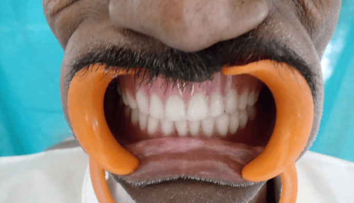
After making the impression with the right technique, the sequential steps of complete denture construction were followed as usual. (Figure – 9).

Discussion
Prosthodontic management of palatal defects has been employed for many years. Ambroise Pare probably was the first to use artificial means to close a palatal defect – as early as the 1500's. The early obturators were used to close congenital rather than acquired defects. The early objectives of treatment were artificial closure of the defect and adequate retention of the artificial closure. The ingenious designs of the early pioneers accomplished these objectives. As time progressed, newer and better concepts of obturators evolved.
The basic objectives of prosthodontic therapy include a comfortable, cosmetically acceptable prosthesis that restores the impaired physiologic activities of speech, deglutition and mastication. The most important objective of prosthodontic care, as DeVan stated, our objective should be “The perpetual preservation of what remains rather than the meticulous restoration of what is missing.” This principle is most important in the treatment of the maxillary defect patient.
The discomfort caused by hard resin presents another problem. Popular maxillofacial prosthodontist, Dr. Albert has almost completely abandoned the use of rigid prosthetic materials for making obturators. Among the synthetized materials, he found silicon-rubber components to be the most effective. They are easy to produce and function well. However, for the permanent obturator, maxillofacial prosthodontist Brown states, “. . . heat-cured methyl methacrylate resin still remains the material of choice for tissue compatibility, environmental resistance and ease of adjustment.”
Summary
A thorough knowledge of what is normal is a must for the dentist to understand the acquired defects with which he interacts. It is important that the clinician become familiar with a technique and master it. It must always be remembered, and patient must be so counselled before treatment, that the clinician can only try to provide alternative means of what the patient has lost, and how successful that alternative will be, depends upon the patient’s ability to accept the defect and to adapt to an alternative environment.
“Not mere survival from disease alone, but a return to a normal functioning life” is a must.


Comments