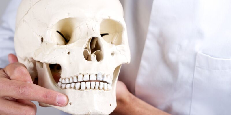
As a significant finding for oral surgery, a recent study has shed light on the relationship between the surface morphology of mandibular third molar roots and the risk of mental nerve paresthesia, a serious postoperative complication.
The study, conducted using dental cone beam computed tomography (CBCT), examined the root morphology of mandibular third molars in 1216 patients who had undergone extractions.
The research, led by a team of dental experts, aimed to fill a critical gap in existing knowledge by investigating the factors contributing to mental nerve paresthesia following mandibular third molar extraction. Surprisingly, no previous study had explored the correlation between root morphology and the likelihood of this complication.
The study focused on 1534 teeth in 791 patients who had undergone CBCT before surgery. Various factors were analyzed: age, complete or incomplete formation of mandibular third molar roots, periodontal ligament atrophy, hypercementosis, and mandibular canal deformation.
One of the key findings was that mandibular third molar root formation is typically completed between the ages of 19 and 30 years. Moreover, the study identified two significant risk factors for mental nerve paresthesia: complete formation of mandibular third molar roots (P = 0.002) and deformation of the mandibular canal (P < 0.001).
These findings imply that the risk of mental nerve paresthesia can be mitigated by performing third molar extractions before the complete formation of the roots.
This insight provides a valuable opportunity for dentists to proactively manage and reduce the occurrence of this serious postoperative complication. Understanding the risk factors associated with mental nerve paresthesia allows us to tailor our approach to mandibular third molar extractions, ensuring the best possible outcomes for our patients.
- Source: DOI: 10.1016/j.ijom.2023.12.008


Comments