
Dentists love to flaunt the curves of treated root canals. With advancements in endodontics, it has become easier to reach normally inaccessible areas in a root canal, helping the clinician to achieve 3D cleaning and obturation.


After the development of new instruments and techniques, use of NiTi rotary instruments has gained popularity; not everything is without its own pros and cons though.

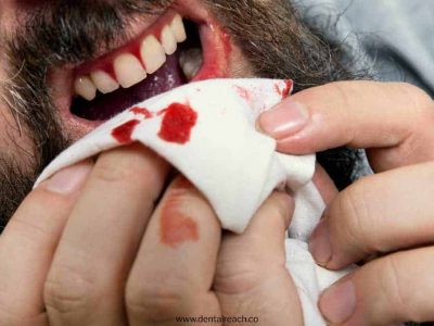
One of the most dreaded nightmares of any clinician is broken instruments in the midst of treatment. NiTi rotary instruments show a high incidence of instrument fracture despite their favourable qualities. Broken instrument not only includes separated files but also applies to a sectioned silver point, a segment of a lentulo, a Gates Glidden drill, a portion of a carrier-based obturator or any other dental material left inside a canal.
Causes
Cinematic movements wrongly applied on instruments. Use of already deformed instruments that can be fractured due to cyclic fatigue.
Treatment
With the advent of newer visualisation techniques, clinicians get an enhanced magnification and illumination by using dental loupes and operating microscopes. It helps in visualising the most coronal aspect of broken instruments and allow them to remove it without perforating the root canal.
There are three options from which a clinician has to choose anxiety attack because of the realisation that the instrument has broken settles down. They are whether to:
- Remove the fractured segment
- Bypass and seal the fragment within root canal
- Go for a true blockage
Factors
There are many factors for the treatment plan:
- Status of pulp
- Root canal infection
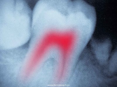
- Root canal anatomy: Higher success rate in cases of anterior teeth with wide and straight canals and for posterior teeth with narrow and curved canals.
- Position of the instrument: The removal of an instrument fragment located in the apical third is particularly complex as compared to middle and coronal thirds. In general, if one third of the overall length of instrument can be exposed, it can be removed.
- Type of broken instrument: It is more difficult to remove NiTi rotary instruments as compared to stainless steel instruments because they generally fracture at a smaller length. They tend to straighten out when they break in a curved canal due to elastic memory. Stainless steel instruments do not break during removal process while NiTi instruments may fracture again and go deeper in the canal presumably due to heat generation.
Amount of damage that can be caused to the remaining tooth structure: Procedure and instruments used to gain access to the broken instrument may increase the risk of perforation of canals.
Along with good technology, a succe
Non surgical method of instrument removal
No attempt to remove the broken instrument should be made unless a proper straight line access to the head of instrument is achieved. It is done step by step by creating a coronal access followed by a radicular access.
Coronal and radicular access
High speed, friction grip, surgical length burs are used to gain straight line access to all canal orifices.Clinical experience suggests that most of the broken files separate towards their terminal extents between D3, D4 and D5.
A predictable way to create a safe radicular access is to initially use hand files, small to large to create sufficient space to use GG drills. The drills are then used to create a smooth flowing funnel which is largest at the orifice and narrowest at the obstruction.
Creating a staging platform
When an ultrasonic instrument introduced into the pre-enlarged canal does not have enough space lateral to the broken segment, to initiate the trephining process, staging platform is created.
This is made by using GG drills whose maximum cross-sectional diameter is slightly larger than the visualized instrument.
Techniques for removal
- Ultrasonic techniques
Prior to performing any radicular removing techniques, cotton pellets should be placed over other open orifices to prevent the re-entry of fragment into another canal. The tip of ultrasonic instrument is placed in intimate contact against the obstruction and typically activated at the lowest power setting that will accomplish the task.
These techniques do not usually generate much heat so procedures can be performed dry to assist visualization. The ultrasonic vibrations help loosen the broken fragment which may sometimes jump out by itself by a slight wedging action.
In case where an instrument is deep inside, a larger length, smaller diameter tip can be used. If it is still deeper, titanium instruments which provide safety due to its smooth cutting action should be used.
- Microtube removal methods
There are several microtube removal methods that are designed to mechanically remove an intracanal obstruction like a broken file. But it requires excessive removal of dentin which may dangerously weaken the tooth structure and predisposes to ledge, perforation and fracture.
Following are the examples of microtube removal techniques:
- Lasso and Anchor use an appropriately sized microtube and a wire passed through the tube, looped at one end and passed back through the tube.
- Tube and Glue uses an adhesive such as core paste to bond the obstruction to the microtube.
- Tap and Thread contain five microtubular taps but its use is limited to radicular obstructions that extend coronally to the pulp chamber or to coronal one-third of the root canal.
- Masserann Kit is an old yet very effective method for strong purchase of instrument and its removal but limited to use in large canals of anterior teeth.
- Spinal Tap Needle is used along with its metal insert plunger or a hedstrom file to remove the broken instrument.
- Endo Extractor is a newer recently released instrument system which is able to gain a strong mechanical purchase on a broken instrument but limited to coronal aspect of larger canals.
- Instrument Removal System consists of three colour coded microtubes each with a different diameter.
Surgical method of instrument removal
In cases where non-surgical removal is not possible like an instrument beyond the apical foramen and intentional leaving of the broken instrument might prove to be risky, surgical approach is undertaken.
This procedure is performed under local anesthesia only after thorough history, proper clinical examination and good quality radiographs.
But as an age old proverb goes:
“ PREVENTION IS BETTER THAN CURE”
- Root canal instruments should be examined before and after use to make certain blades are regularly aligned. Too much or too little space is an indication that the instrument has been under strain and may break.
- The degree of torque applied should be in a controlled manner.
- Instruments should be used according to the sequence without skipping any size.
- Debris should be removed between the blades from time to time.
- Instruments should be used with agents used to wet the canal like sodium hypochlorite and chemicals should be used to facilitate cutting where necessary.
References:
- Ruddle, CJ. Nonsurgical endodontic retreatment. J Calif Dent Assoc. 200432:4
- Friedman, S, Stabholz, A. Endodontic retreatment: case selection and technique. Part 1: criteria for case selection. J Endod. 1986;12:28–33.
- Stabholz, A, Friedman, S. Endodontic retreatment: case selection. Part 2: treatment planning for retreatment. J Endod. 1988;14:607–614.
- Ruddle, CJ. Microendodontic nonsurgical retreatment. in: Microscopes in Endodontics, Dent Clin North Am. 41. W.B. Saunders, Philadelphia; 1997:429– Johnson, WT, Leary, JM, Boyer, DB.
- Smith, BJ. Removal of fractured posts using ultrasonic vibration: an in vivo study. J Endod. 2001;27:632–634.
Johnson, WT, Leary, JM, Boyer, DB. Effect of ultrasonic vibration on post removal in extracted human premolar teeth. J Endod. 1996;22:487–488


Comments