– Dr. Urvashi Tanwar


Nothing can replace the natural, but the natural! A sub-gingival tooth fracture does NOT always mean extraction of the fragment/tooth, but rather retaining the natural tooth.


Introduction
Crown fractures of the permanent dentition hold the highest percentage of traumatic injuries in the oral cavity, some of them being complex and involving the pulp. When the trauma causes a subgingival fracture line, the prognosis is considered to be questionable to hopeless at best.
In order to gain a healthy tooth-gingival relationship, placing crown margins supra-gingivally, the endodontic management, impression techniques, followed by restorative and prosthetic procedures-while also keeping the esthetic demands of the patient in mind, often warrant a complex interdisciplinary approach.
Etiology

Heithersay’s Classification
The severity of the fracture in a subgingival direction is the most important factor influencing the treatment plan and can be classified as 1:
- Type 1: Teeth with subgingival fractures, where no portion of the fracture line extend below the level of the attached gingiva (1 to 2 mm).
- Type 2: Teeth with subgingival fractures where a portion of the fracture line extends below the level of the attached gingiva, but does not extend below the level of the alveolar crest (2 to 4 mm).
- Type 3: Teeth with subgingival fractures where a portion of the fracture line extends below the level of the alveolar crest (more than 4 mm).
- Type 4: Teeth with root fractures where the fracture line is within the coronal one-third of the root and totally below the level of the alveolar crest.

Clinical Presentation
- Most patients are asymptomatic, but some may complain of pain when the fragment is partially attached to the periodontal ligament or the fracture is in close approximation to the pulp, and while eating/chewing due to movement of the coronal fragment.
- Delayed detection and treatment would cause serious inflammatory changes of the pulp and gingiva, clinical attachment loss and bone loss manifesting clinically as gingival bleeding and deep periodontal pockets.
Management of Sub-gingival Fractures in Anterior Teeth
Assessment: A thorough assessment of the hard tissue (fracture line) as well as soft tissue (lacerations) surrounding the fracture site is of prime importance.
Before starting treatment, the dentist must evaluate:

Investigations
- Radiographs do not offer much help as the fracture line is usually parallel to the central X- Ray beam.
- For detection of fracture site, magnification/dyes can be used.
- CBCT: Helps in assessment of level of fractures whereas the reconstructed axial views were effective in detection of fractures. Combining the clinical and radiographic data play a crucial role in treatment planning.
Restorative Challenges Encountered
- Diminished/absent coronal ferrule
- Compromised biological width
- Difficulties in access and isolation
Through the years, there have been mainly two methods of gaining coronal ferrule while re- establishing biological width:
- Surgical Crown Lengthening and
- Orthodontic Extrusion.
Fractures treated at the earliest (24 hours) have the best prognosis.
Treatment Options & Considerations in Anterior Teeth
Treatment options available are:
- Removal of the coronal fragment with subsequent restoration above the gingival level.2
- Removal of the coronal fragment supplemented by gingivectomy and osteotomy and subsequent restoration with the crown and nucleus. 3
- Removal of the coronal fragment, raising of a gingival flap, immediate endodontic treatment and fragment bonding.4
- Removal of the coronal fragment and immediate extrusion of the root by surgical ororthodontic procedures.5
- Extraction when the fracture is more than one-third of the root.6

A. Endodontic Perspective:
Root canal treatment is almost always indicated before starting with marginal acquisition due to communication of fracture line with the chamber or for ease of future restorative/prosthetic rehabilitative procedures.
Following appropriate pulpal therapy, soft tissue management of increasing the crown length can be done through surgical or orthodontic procedures.
Crown Lengthening: A procedure which aims to increase the supragingival tooth structure for esthetic and restorative purposes. It can be done either surgically or orthodontically. To restore a damaged tooth by crown lengthening, the coronal extent of the remaining tooth structure should have a minimum length of 3.5-4 mm from the alveolar crest.7
B. Periodontal Perspective:
- Surgical crown lengthening can be done by gingivectomy or an apically repositioned flap with or without osseous recontouring.
- Gingivectomy will suffice if only 1-2 mm of crown height needs to be increased and if the tooth has sufficient width of attached gingiva with a probing depth of 4 mm.8,9
- Apical repositioning of the flap with bone recontouring is done when more amount of crown has to be exposed.

C. Orthodontic Perspective
Introduced by Heithersay in 1982, extrusion of a tooth is done by applying traction to the periodontal ligament stimulating marginal apposition of crestal bone and allowing en masse vertical movement of the dento- gingival apparatus. It can be:
- Slow: Gingiva, periodontal ligament and alveolar bone migrate coronally along with the tooth under low intensity extrusive forces.
- Rapid: When stronger traction forces are exerted, coronal migration of the
tissues supporting the tooth is less pronounced because the rapid movement exceeds their capacity for physiologic adaptation. The alveolar bone is left behind temporarily and to prevent the bone from moving coronally, circumferential fiberotomy is performed.

Maximum force for a slow movement should not exceed 30 g, whereas rapid extrusions are accomplished with forces higher than 50 g.

COMBINATION TECHNIQUE: Taking into account the case dynamics, a combination technique incorporating both orthodontic as well as periodontal procedures can also be carried out to overcome the limitations of either technique, but get the best of both.
D. Restorative/Prosthetic Perspective
- Immediate reattachment of fragment is a conservative temporary treatment option, provided isolation is achievable.
- The contour of the crowns must not be exaggerated to compensate for the constricted cervical dimension of the root post extrusion.
- Embrasures should not be filled to prevent an over- contour, as that can affect the marginal periodontium. 10
- Biologically oriented preparation techniques (BOPT) prepared with feather-edge preparation can be tried following surgical extrusion.
- Newer post systems has caused major developments in coronal as well as intra-radicular rehabilitation allowing quick placement and completion of prosthetics in young permanent teeth.

Comprehensive Treatment Plan for Heithersay’s Classification 1

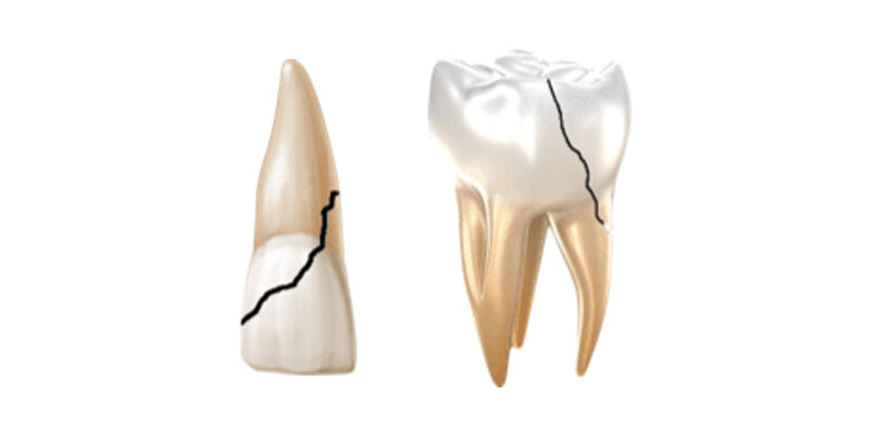
Management of Sub-gingival Fractures in Posterior Teeth
Cracks/Fractures of posterior teeth extended sub-gingivally can be mainly of 2 types: (according to AAE)


Treatment Options & Considerations in Posterior Teeth
The treatment options depend on mainly whether it is a fractured cusp or a cracked tooth. Hence, understanding the difference between them is of utmost importance. Final treatment also depends on other considerations like:

Future Trends
- Mini-implant guided extrusion is slowly gaining popularity. Less invasive procedures like Corticision 11, Piezocision 12 and Piezopuncture 13 promoting accelerated tooth movement have been reported in the literature.
- Microscope helps in the conservative management yielding more predictable outcomes.
- Diode lasers have made soft tissue management of marginal elevation easier offering excellent hemorrhage control.
- Minimalistic management by deep margin elevation (DME) can be attempted in cases where pulp is not involved, followed by replacement of the fractured fragment with direct/indirect methods.
- Advances in bonding strategies as well as development of newer bioactive restorative materials opens opportunities to conservatively restore fragments in the least invasive way possible.
To Summarise


Conclusion
Interdisciplinary management of sub-gingivally fractured teeth mainly depends on accurate assessment of all factors that will affect the prognosis of the tooth in the oral cavity. Attempts must be made to achieve a supra-gingival crown margin through the most conservative and least invasive methods as dictated by the case. Newer techniques and restorative materials must be tried when indicated to be minimally invasive while treating such cases.
Henry Ford said, coming together is a beginning, staying together is progress, but working together is SUCCESS!
References
- Heithersay GS, Moule AJ. Anterior subgingival fractures : a review of treatment alternatives. Aust Dent J 1982; 27(6):368–76.
- Roeters J, Bresser JP. The combination of a surgical and adhesive restorative approach to treat a deep crown/root fracture: a case report. Quintessence Int 2002;33:174^9.
- Andreasen JO, Andreasen FM. Traumatismo Denta ̈ rio: solucËo¬ es cl| ̈nicas. Sa¬ o Paulo: Panamericana;1991.168pp.
- MeloLL.TraumatismoDenta ̈rio,1stedn.Sa¬oPaulo:Artes Me ̈ dicas;1998.
- BondemarkL,HallonstenA.Attractivemagnetsforortho- dontic extrusion of crown^root fractured teeth. Am J Orthod Dentofacial Orthop1997;112(2):187^93.
- Magini RS, Censi JC, Bianchini MA. Reimplante Intencional para Tratamento de Fissura Longitudinal: relato cl| ̈ nico apo ̈ sacompan hamento de um ano. Rev Bras Odont 1997;54:297^302.
- Ayush Razdan Singh, Ruchita Verma. Crown lengthening versus forcederuption. Orthodontic Journal of Nepal .2011; 1(1) : 52- 55.
- Liudvikas Planciunas, Alina Puriene, Grazina Mackeviciene. Surgical lengthening of the clinical tooth crown. Stomatologija, Baltic Dental and Maxiollofacial Journal. 2006; 8: 88-95.
- Marianne Ong, Shin-Chang Tseng, Hom-Lay wang. Crown Lengthening Revisited. Clinical Advances in Periodontics. 2011; 1(3).
- Cronin RJ, Wardle WL. Prosthodontic management of vertical root extrusion. J Prosthet Dent 1981; 46(5):498–504.
- Kim SJ, Park YG, Kang SG. Effects of Corticision on paradental remodeling in orthodontic tooth movement. Angle Orthod 2009; 79(2):284-91.
- Keser EI, Dibart S. Piezocision assisted invisalign treatment. CompendContin Edu Dent 2011; 32(46-48): 50-1.
- Vercelloti T, Podesta A. Orthodontic microsurgery: A new surgicallyguided technique for dental movement. Int J Periodontics RestorativeDentistry 2007; 27: 325-331
- Google Images/Images from Orthodontic Extrusion: Periodontal Considerations and Applications Normand, Bach et al.


Comments