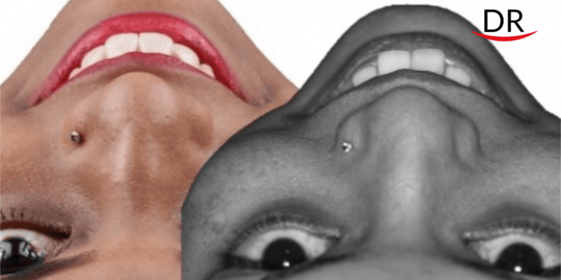
Dr. Smita Kole is a master in composite & aesthetic dentistry (Japan). She maintains a private practice at Solapur, India since 15 years. She regularly keeps herself updated by participating in courses and CDE programs. She is a recipient of multiple dental awards. She is a coveted speaker at the upcoming DRDCA 202 International.
Co authors – Dr. Vaishali Jindam (BDS), Dr. Sushmita Pukale (BDS), Dr. Nonie Nankani (BDS).
Case Report
A 23-year-old female patient presented to our practice with a concern of a discolored anterior tooth and also complained of proclination in her anterior teeth even after orthodontic correction.
On examination
- Discolored 11 with no pain, sensitivity and tenderness.
- Proclined maxillary anterior teeth and spacing between 21 & 22.
- Low frenum attachment.

Radiographic examination was done for 11 which indicated a periodical radiolucency.
After the complete examination, patient was explained about the various treatment options, the number of visits required and the finances involved. She was also informed about her gingival and frenum positioning and a need to undergo additional procedures of laser assisted frenectomy and gingivoplasty to correct the gingival zenith. Initially patient was reluctant about the entire procedure, however after proper counselling with visual aids, agreed to go ahead with the treatment.
Treatment plan
- Complete oral prophylaxis
- Root canal procedure for 11 followed by a zirconia layered with ceramic crown
- Frenectomy and gingivectomy for correction of gingival zenith
- Lithium di-silicate veneers with 12,21,22 and added enameloplasty with 13
Procedure Summary
- According to the plan, a thorough oral prophylaxis was done.
- The discolored tooth (11) shade (A3) was recorded for further comparison using VITA classic shade guide. The adjacent natural tooth shade was also recorded (B1).

- Impressions and wax mockups were made, ready to give the patient an idea of the end result through the trial run. Patient was quite impressed with the result and decided to go forward with the treatment.

- As planned 11 was treated endodontically.
- The laser assisted correction of the gingival zenith and low frenum attachment was performed.

- Tooth preparation was done for 11, 12, 21, 22, while doing the preparation there was a need for an intentional RCT in 21 to compensate for the proclination.
- This was followed up giving a set of temporaries on the prepared teeth using Protemp 4. Patient was very happy with the idea of what the final outcome would be.
- As planned, the Zirconia layered with ceramic crown for 11 and Lithium Disilicate veneers for 12, 21, and 22 were received from the lab, which were then tried out intraorally.

- After a satisfactory trial, the prosthesis were made ready for cementation.
- Zirconia crown was cleaned with Ivoclean (Ivoclar) for 20 seconds and rinsed thoroughly with water. Silane coupling agent (MonobondPlus, Ivoclar) was applied and allowed to air dry for 60 seconds, followed by application of a dual cure bonding agent (Palifique Universal bond, Tokuyama).
- The lithium disilicate veneers were etched with 5% hydrofluoric acid for 20 seconds (Ceramic etching gel, Ivoclar Vivodent) followed by the silane coupling agent (Monobond Plus, Ivoclar) and a dual cure bonding agent (Palifique Universal Bonding agent, Tokuyama).

- Meanwhile rubber dam isolation was done; etching was carried out with 37% phosphoric acid for 15-20 seconds; teeth were air dried. A Dual cure bond (Palifique Universal Bonding agent, Tokuyama) was applied. The prosthesis was cemented intraorally using a dual cure adhesive resin cement (RelyX Ultimate adhesive Resin Cement, 3M). The excess was removed after a 5 second tack cure, after which the final curing was completed.

- Complete 1min light curing done through KY jelly for oxygen inhibition layer.
- To finish it off, enameloplasy was done with 13 so that it could resemble 23.

- We also planned for composite buildups on 14 and 24, but patient was already satisfied with the results.


Comments