– Dr Mohini Daultani


Introduction
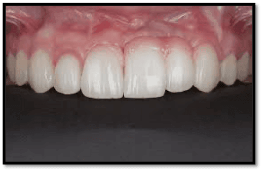
The treatment of localised edentulous area with ridge defects poses a challenging task for the dentist for prosthetic rehabilitation. (Fig 1a & Fig 1b)


Etiology of Loss of Alveolar Bone – Tissue defects can be caused because of many reasons, like:

Siebert’s Classification – Soft tissue defects can be classified as:

Many different techniques are available for predictable bone augmentation. A variety of factors such as quality and quantity of existing contiguous hard and soft tissues, systemic conditions and economic status of the patient play a important role in treatment planning, clinical outcome and prognosis. (Fig 2a & Fig 2b)


Tissue augmentation– Surgical techniques to enhance alveolar bone volume include:

This case report presents the surgical management of Seibert’s class I defect (bucco-lingual) using connective tissue graft by the envelope flap technique.
Case History

Patient reported to the clinic with a chief complaint of missing teeth. On intraoral examination, 22 and 23 were found to be missing with a soft tissue defect bucco-lingually. Radiological examination was done by means of an IOPA with 22,23.
The diagnosis was – Seibert’s Class I defect with 22,23. The soft tissue grafting technique was planned using connective tissue graft obtained from palate. The flap technique planned was envelope- flap technique under local anesthesia.
SURGICAL STEPS
Fig 3: Preoperative view with 22,23 with Seibert Class I defect.

Fig 4: Incision placed at crestal area

Fig 5: Flap reflection -split thickness flap

Fig 6: Template placed at donor site

Fig 7: Incision placed at donor site

Fig 8: Connective tissue graft obtained

Fig 9: Sutures placed at the donor site

Fig 10: Connective tissue graft placed at recipient site

Fig 11: Flap closed at recipient site

Fig 12: Sutures placed at recipient site


Fig 13: COE pack placed at recipient site
Postoperative instructions were given.

Fig 14: Post operative View-1 month follow-up

Fig 15: Preoperative & Postoperative


Prosthesis was planned only after gain in bucco-lingual defect fill.
Fig 16: Pink ceramics


Discussion
In today's practice, patients with normal skeletal pattern who have lost a substantial degree of their original osseous dimensions due to tooth loss or trauma, are much more prevalent. Alveolar ridge defects are common and pose a significant problem in dental treatment and rehabilitation. Therefore, multi-disciplinary approach is most applicable to the many problems involved in the successful reconstruction of localized defects that exist within the alveolar ridge.
The various treatment modalities are:
Sub-epithelial connective tissue graft – We chose to go for a sub epithelial connective tissue graft over other options because if its various advantages like:

Tissue augmentation– A good prosthesis ultimately depends on good underlying tissue. This objective can be achieved if teeth are extracted in an atraumatic manner and appropriate bone graft material are placed into the sockets (Socket preservation) to prevent the eventual collapse of the ridge (Fig 17).

Guided tissue regeneration procedures also may be used to prevent collapse within the ridge or augment an existing defect (Fig 18).

Sequential step wise reconstruction is essential for long term success of rehabilitation.
Prosthetic options– For improvement of esthetics in alveolar ridge deformity, there are various prosthetic and surgical options available.Gingival (pink) ceramic or long pontic design can be used in the cervical area to improve the esthetics in such cases, just like it was used in this one.
Conclusion
Various techniques and materials for alveolar ridge augmentation are available today, but it is most appropriate to use an multi-disciplinary approach when a treatment plan is being developed for bone augmentation cases. It is thus evident that for prosthetic restorations with optimal function and esthetics, an interdisciplinary team approach is mandatory. Important factors in achieving long term success for rehabilitation include:
- proper examination
- diagnosis
- treatment planning
- careful execution of the surgical procedures and
- postoperative follow-up.
References
- Manjunath N, Arjun M R. An ounce of prevention is worth a pound of cure: A review on ridge augmentation. J Interdiscip Dentistry 2015;5:97-104
- Jin QM, Anusaksathien O, Webb SA, Rutherford RB, Giannobile WV. Gene therapy of bone morphogenetic protein for periodontal tissue engineering. J Periodontol 2003;74:202-13
- Orth CF. A modification of the connective tissue graft procedure for the treatment of type II and type III ridge deformities. Int J Periodontics Restorative Dent 1996;16:266-77.
- Summers RB. A new concept in maxillary implant surgery: The osteotome technique. Compendium 1994;15:152, 154-6, 158
- Seibert JS, Louis JV. Soft tissue ridge augmentation utilizing a combination onlay-interpositional graft procedure: A case report. Int J Periodontics Restorative Dent 1996;16:310-21


Comments