This article is the winning entry of the DRDCA 2020 Article Contest (First Position). Congratulations to the author Dr. Mohini Daultani!


Abstract
Dark or black colored gingiva is an esthetic concern especially in subjects with high lip line or gummy smile. Gingival depigmentation procedure is a type of perioplastic surgery where the gingival epithelium is excised. Repigmentation is spontaneous process and has been attributed to the activity and migration of melanocytic cells from surrounding areas. Here we present a case report of gingival melanin repigmentation after diode laser depigmentation with a three years followup.

Key-words: Depigmentation, Diode laser, Melanin pigmentation, Repigmentation.
Key Messages: This is a case report of laser depigmentation procedure with a three year follow-up to evaluate melanin repigmentation. In the present case report, we observed spots of melanin repigmentation at the end of one year, which turned more diffused by the end of three years. Long term follow up of such patients should be carried out to determine the timeframe of complete recurrence of repigmentation.
INTRODUCTION
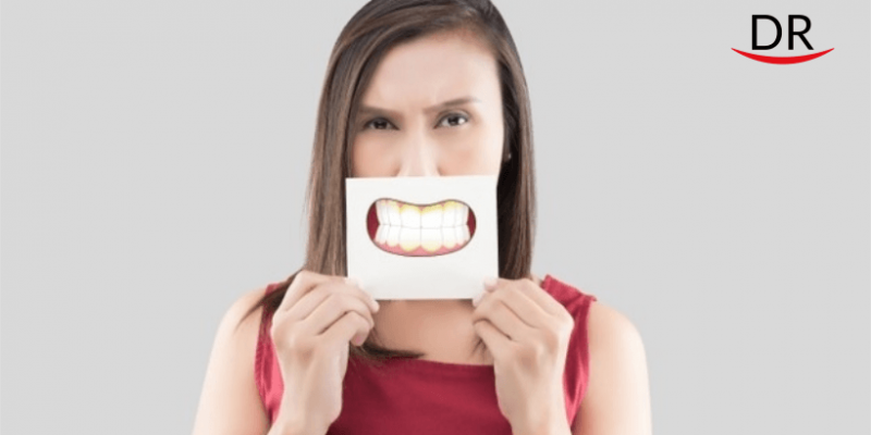
The beauty of a smile is not only influenced by how the teeth and lips look, but also by the way gingival tissue appears. Dark pigmentation of the gums occurs due to excessive melanin deposition in the gingival epithelium. Melanin pigmentation of the gingiva is a physiologic process that occurs in all ethnicities [1]. The prevalence of melanin pigmentation varies between 0% and 89% in different populations with regard to ethnic factors and tobacco usage [2]. Gingival hyperpigmentation is termed as physiological or racial pigmentation, as it occurs as a genetic trait in some populations [3]. Gingival hyperpigmentation in patients with gummy smile or excessive gingival display could pose as an esthetic problem.
Gingival depigmentation is a periodontal plastic procedure carried out by various techniques like conventional scalpel technique, gingivectomy, free gingival graft, bur abrasion, cryotherapy, electrosurgery, and laser therapy. The choice of the surgical technique for depigmentation is based on individual preferences and clinician’s experience.
The clinical reappearance of melanin pigment following a period of depigmentation is referred as “repigmentation”. Post-surgical repigmentation of gingiva has been reported in the literature. Repigmentation is spontaneous process and has been attributed to the activity and migration of melanocytic cells from surrounding areas.
Melanin repigmentation has been observed in all the surgical depigmentation procedures [4]. There is a varying rate of repigmentation after surgical depigmentation procedures which range from 1 month to 8 years.
We present a case report of gingival melanin repigmentation after laser depigmentation with a three years followup.
CASE REPORT
A 22-year-old female patient complaining of heavily pigmented gums (Figure 1) visited department of Periodontology, School of Dental Sciences, Karad.

On examination, the patient was systemically healthy but had a gummy smile with darkly pigmented gingiva. The dark color of gingiva was due to physiologic excessive melanin pigmentation seen in Asian ethnic origin. Dummett Gupta oral pigmentation index (DOP) [5] was used to grade the level of gingival hyper pigmentation (Scoring criteria: 1, No clinical pigmentation; 2, Mild clinical pigmentation; 3, Moderate clinical pigmentation; 4, Heavy clinical pigmentation). DOP score of three (moderate clinical pigmentation) was observed in our patient. The pigmentation was esthetically displeasing and hence gingival depigmentation procedure to lighten the color of gingiva was planned. The subject was educated about her condition and informed about the various techniques available for depigmentation procedure. It was decided to use diode laser with wavelength 980 nm to carry out gingival depigmentation procedure after obtaining an informed consent.
All the necessary laser operatory precautions were followed during the procedure. A semiconductor diode surgical laser unit (Photon Plus; Zolar Tech Technology & MFG Co Inc, Canada, wavelength 980 nm, power 10 W) was used for depigmentation. Local anesthesia (LOX 2%, Neon laboratories Ltd, Andheri Mumbai) was administered through infiltration anesthesia in the operating area i.e. maxillary anterior gingiva up to the distal line angle of canine on both right and left sides. The semiconductor diode laser was emitted in gated-pulsed mode, and was operated in a contact method using a flexible fiber optic handpiece (Table 1).

Laser ablation was started from the mucogingival junction toward the free gingival margin, including papillae. The laser tip was moved using light brushing strokes to prevent heating of the tissues. The procedure was performed in contact mode in cervico-apical direction on pigmented areas (Figure 2). The area was irrigated using saline and the charred epithelium over the tissue was removed with saline-soaked gauze.Laser ablation of the anterior labial sextant of upper and lower arch was completed within 30 minutes. Periodontal dressing (COE-PAK, GC America Inc, ALSIP, IL, USA) was applied over the wound surface. No pain or bleeding complication was observed during and after the procedure.

Patient was instructed to avoid eating hot and spicy foods for the first 24 hours and was discharged from the dental office to resume normal daily activity. Patient was recalled after seven days for removal of periodontal pack. Healing was satisfactory at the end of one month with pink appearance of gingiva with Dummett Gupta score 0, resulting in a significant improvement in an esthetic appearance (Figure 3).

The case was followed for three years after laser depigmentation procedure. Evaluation of melanin repigmentation was carried out every six months by Dummett Gupta score. At the end of one year, there were spots of melanin repigmentation observed in the attached gingiva. At the end of second year the spots had expanded and appeared as diffuse gingival pigmentation involving attached gingiva and interdental papilla. At the end of three years the Dummett Gupta score had increased to 2 (mild clinical pigmentation) which was observed in attached gingiva, interdental papilla, and marginal gingiva (Figure 4). Melanin repigmentation was less prominent post operatively as compared to pre operative photograph.

DISCUSSION
Darkly pigmented gingival tissue forces the patients to seek cosmetic treatment. Several treatment modalities have been suggested and presented in the literature ranging from a simple slicing method to free gingival grafts or "push back" operation. Many easy, simple and effective techniques are described, which gives desired results. Melanin pigment recurrence has been documented to occur, following all the surgical depigmentation procedures. The epithelium melanin unit is formed by the melanocytes and keratinocytes. There is little information on the behavior of melanocytes after surgical injury. A study carried out by Oswaldo et al and Kon et al showed that gingival surgical procedures performed solely for cosmetic reasons, offer no permanent results [6].
The large variation in time of repigmentation may be related to the technique used and the race of the patient. Repigmentation may also be attributed to melanocytes, which are left during surgery as stated by Ginwalla et al. These may become activated and start synthesizing melanin. Atsawasuwan et al. reported the use of Nd: YAG laser for gingival depigmentation in four cases. The Nd: YAG laser was set at 6 watts, 60 millijoules per pulse, and 100 pulses per second. They found no recurrence of pigmentation during the follow up period of 12 months. The authors concluded that Nd : YAG laser had shown to be a good option for gingival depigmentation, and caution must be exercised in delicate areas near marginal gingiva while using Nd : YAG laser [7] . Perlmutter and Tal reported the case of one patient in whom gingival repigmentation occurred after 7 years of gingival laser depigmentation [8].
Dummett and Bolden observed partial recurrence of repigmentation in six out of eight patients after gingivectomy at 4 months [9]. Nakamura et al described depigmentation with CO2 laser in ten patients. No repigmentation was seen in first year, but four patients showed repigmentation by 24 months [10].
Spontaneous repigmentation has been shown to occur and the mechanism suggested is that, the melanocytes from the adjacent pigmented areas proliferate and migrate into the depigmented areas. Further research is required on repigmentation for studying the factors affecting rate and length of time required for recurrence of pigmentation. Research should also focus on finding a solution for preventing the recurrence and till then, repeated depigmentation should be carried out to eliminate the unsightly pigmented gingiva.
CONCLUSION
The application of diode laser (980nm) for melanin depigmentation is a safe and effective tool with no post-surgical complications. In the present case report, we observed spots of melanin repigmentation at the end of one year, which turned more diffused by the end of three years. Long term follow-up of such patients should be carried out to determine the timeframe of complete recurrence of repigmentation.
REFERENCES
[1] Dummett CO. Clinical observations on pigment variations in healthy oral tissues of the Negro. J Dent Res 1945;24(1):7-13.
[2] Dummett CO, Barens G. Oromucosal pigmentation: an updated literary review. J Periodontol. 1971;42:726-36.
[3] Prinz H. Pigmentation of the oral mucous membrane. Dental Cosmos 1932;72:554-61.
[4] Suragimath G, Lohana MH, Varma S. A split mouth randomized clinical comparative study to evaluate the efficacy of gingival depigmentation procedure using conventional scalpel technique or diode laser. J Lasers Med Sci. 2016;7:227-32.
[5] Dummett CO. Physiologic pigmentation of the oral and cutaneous tissues in the negro. J Dent Res. 1946;25:421–32
[6] Kathariya R, Pradeep AR. Split mouth de-epithelization techniques for gingival depigmentation: A case series and review of literature. J Indian Soc Periodontol 2011;15:161-8.
[7] Atasawasuwan P, Greethong K, Nimmanon V. Treatment of gingival hyperpigmentation for esthetic purposes by Nd:YAG lasers; Report of 4 cases J Periodontol 2000;71:315-21
[8] Perlmutter S, Tal H. Repigmentation of gingiva following surgical injury. J Periodontol 1986;57:48-50.
[9] Dummett CO, Bolden TE. Postsurgical clinical repigmentation of the gingiva . Oral Surg, Oral Med, Oral Pathol 1963;16:353-57.
[10] Nakamura Y, Hossain M, Hirayama K, Matsumoto K. A clinical study on the removal of gingival melanin pigmentation with the CO2 laser. Laser Surg Med 1999;25:140-47
Disclaimer – The views and opinions expressed in this article are that of the author alone and does not necessarily reflect the official policy of DentalReach. DentalReach does not endorse, promote or associate with this product and this article is meant for informative purposes only.


Comments