
INTRODUCTION
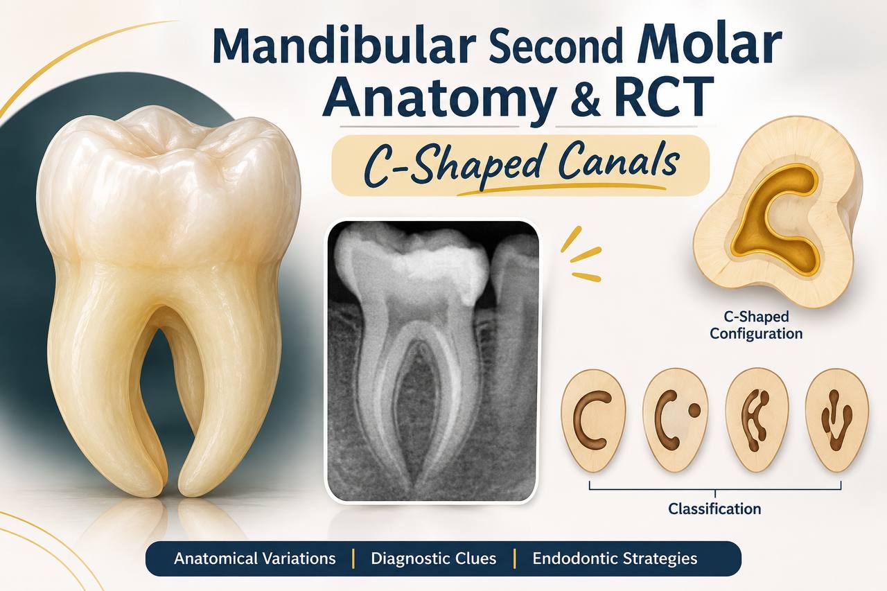
Successful root canal treatment depends on understanding the internal anatomy of the tooth and disinfecting the entire root canal system before obturation. Although mandibular second molars commonly present with two roots and separate mesial and distal canals, variations such as fused roots and C-shaped canal systems may occur. A C-shaped configuration is frequently associated with fusion of the roots and a longitudinal groove on the external root surface. In cross-section, the canal may resemble the letter “C,” but its shape can change considerably from the orifice to the apical third.
Melton and colleagues described a classification based on canal cross-sectional form, while Fan and colleagues later proposed a commonly used system describing the configuration at different levels of the root. The major patterns include a continuous C-shaped lumen (C1), a semicolon-shaped lumen (C2), two or three separate canals (C3), a single round or oval canal (C4), and no visible canal lumen (C5, usually near the apex). These configurations are not static: one tooth may show different patterns at different root levels. Consequently, a single access view or radiograph may not reveal the full complexity.
The clinical significance is substantial. Thin fins and isthmuses may remain untouched by rotary instruments, while aggressive circumferential enlargement can unnecessarily weaken already thin radicular dentine. Irrigation and sonic or ultrasonic activation along with appropriate working-length control and a three-dimensional obturation strategy therefore have a central role in managing these cases.
CASE PRESENTATION
PATIENT HISTORY AND CLINICAL FINDINGS
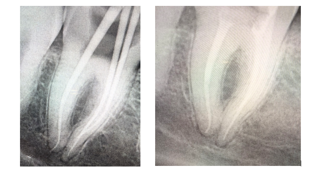
A 27-year-old patient reported pain in the lower right posterior region for approximately since 10 days. Clinical examination revealed deep caries involving mandibular right second molar (FDI tooth 47) where caries was approaching pulp. Based on the history- patient was having aggrevating pain in night whereas pain subsided only after taking medicaons for 2-3 hours. Along with radiograph assessment , the pulpal diagnosis was symptomatic irreversible pulpitis. The periapical status revealed no periapical infection or abscess present with slight findings of PDL widening. Whereas the sensibility-test responses was negative and tooth was non vital due to deep caries and restorability assessment states that mesiobuccal and mesiolingual wall was completely fractured.
RADIOGRAPHIC ASSESSMENT
The submitted radiographs document the root canal treatment procedures from master con to final obturation of tooth 47 . The images suggest a complex canal configuration and demonstrate radiopaque root fillings extending through the visible canal paths suggesting c shaped canals. As these are two-dimensional projections, they cannot independently establish the exact C-shaped subtype or exclude additional fins, isthmuses or canal communications.
ENDODONTIC CONSIDERATIONS AND TREATMENT APPROACH
Following diagnosis and consent, treatment planning should prioritize asepsis, preservation of tooth structure and disinfection of the full canal system. Rubber-dam isolation is recommended for endodontic treatment. After access preparation, the chamber floor should be inspected under magnification and the canal orifices explored gently with small hand files. Tactile feedback, electronic apex location and appropriately angled radiographs can assist working-length determination. In a suspected C-shaped system, the clinician should avoid forcing instruments into narrow fins or attempting to convert the entire C-shaped space into a circular preparation.
Free for members
Keep reading — it's free
The rest of this article is open to DentalReach members. Create a free account, or sign in, and every article on the site opens in full.