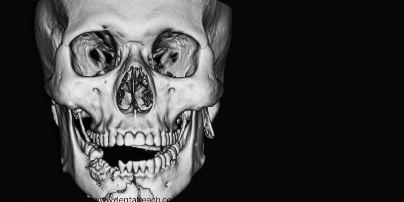
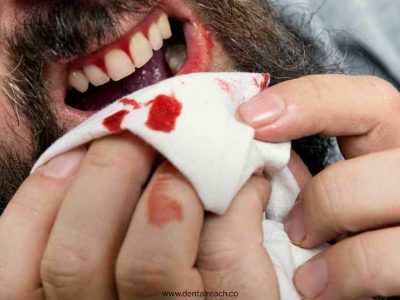
TRAUMATIC INJURIES OF TEETH
CONCUSSION
- Concussion refers to vascular structures at the tooth apex and periodontal ligament resulting in inflammatory edema
- No displacement, only minimal loosening of tooth occurs
- May result in mild avulsion of the tooth from its socket causing occlusal surface to make premature contact with an opposing tooth
Clinical features
- Tenderness on gentle horizontal or vertical percussion
- Tooth sensitive to biting forces
- Patients usually try to modify occlusion to avoid traumatized tooth
Management
- Soft diet
- Relief of occlusal interferences
- Flexible splinting
- Periodic monitoring with repeated vitality testing and radiographs
Prognosis
- Pulp necrosis
- Root resorption is very rare
LUXATION
- Dislocation of the tooth from its socket after severing of the periodontal attachment
- Usually two or more teeth involved
- Teeth mostly affected: deciduous and permanent maxillary incisors
- Mandibular teeth seldom affected
- Vitality testing: temporarily decreased or undetectable
- Vitality may return after weeks or several months
- Depending on magnitude and direction of traumatic force
- Subluxation
- Extrusive luxation
- Lateral luxation
- Intrusive luxation
SUBLUXATION
Subluxation denotes an injury to supporting structures of the tooth that results in abnormal loosening of the tooth without frank dislocation.
Clinical features
- Teeth are in normal location or limited elevation of tooth from its socket
- Abnormally mobile
- Extravasated blood emanating from gingival crevice depicts PDL damage
- Tenderness to percussion and masticatory forces
EXTRUSION
- Partial displacement of a tooth out of its socket
- Often found in deciduous teeth
Clinical features:
- Tooth appears elongated
- Usually displaced palatally
- Bleeding from gingival sulcus
- Mobile
LATERAL LUXATION
Movement of tooth in a direction other than intrusive or extrusive displacement
Clinical features
- Comminution or crushing of alveolar process accompany tooth dislocation
- Movement direction depends on:
- Orientation and magnitude of the force
- Root shape
- Tooth may be pushed through buccal or less commonly lingual cortical plate
- Root apex palpable insulcus area
Management (Subluxation, Extrusion, Lateral luxation)
- Restoring teeth to normal position by digital pressure under LA
- Comminuted pieces of alveolar bone to be repositioned by digital pressure
- Removal of occlusal interferences if necessary
- Immobilization for 2-3 weeks using flexible splints
- Root canal therapy prior to splint removal
- Extraction of the traumatized teeth should be the last resort
- Periodic follow up clinically and radiographically
Prognosis
- Pulp necrosis: Open apex -9%, Closed apex -55%
- Chances of surface resorption
- Inflammatory resorption can be seen in association with pulp necrosis
- Due to compression to the PDL, both inflammatory and replacement resorption may occur
INTRUSION
- Displacement of tooth into alveolar process
- Comminution or crushing of alveolar process accompany tooth dislocation
- Often seen with deciduous dentition, less in permanent dentition
Clinical features
- Reduced height of clinical crown
- Gingival bleeding evident
- High metallic sound on percussion
- Maxillary incisors may be intruded into the alveolar process
- Damage to adjacent teeth especially underlying permanent teeth
Management for intrusion:
Depends entirely upon the stage of root development
Immature root formation
- Spontaneous eruption can be anticipated
- Luxation of tooth slightly with the forceps done if no signs of re-eruption after 10 days
- Pulpal healing is monitored during the period of re-eruption at 3, 4, 6 weeks after injury
- In case of negative response of the pulp or periapical radiolucency
- Endodontic therapy with calcium hydroxide dressing is done
Completed root development
- Spontaneous re-eruption is unpredictable
- Orthodontic extrusion is indicated over a period of 2-3 weeks
- Prophylactic endodontic therapy is indicated as frequency of pulp necrosis
Prognosis
- Pulpal necrosis-Open apex-63%, Closed apex –100%
- External surface, inflammatory and replacement resorption are very frequent findings, especially in teeth with complete root development
- Severe complication can be seen as late as 5-10 years after trauma
AVULSION
- Complete displacement of a tooth from the alveolar process
- Can occur due to direct or indirect trauma
Clinical features
- Seen in relatively younger age group
- Maxillary central incisors-most commonly avulsed teeth in both dentitions
- Affects single tooth mostly
- Socket is found empty or filled with coagulum
- Lip laceration
- Fracture of alveolar process may occur
Management
- If avulsed tooth is not found clinically or radiologically, chest or abdominal radiograph to locate it
- Reimplantation of permanent teeth. The prognosis depends on:
- Condition of tooth
- Time out of socket
- Viability of residual PDL fibres
- Splinting
- Endodontic therapy after reimplantation
- Follow up
Bonus: Download our monthly e-bulletin!Click here to get it
DISCLAIMER : “Views expressed above are the author’s own.”


Your support is highly appreciated. Glad that the information was useful.
Hi nikhat.hows u dear.
Wer u vanished.