The removal of mandibular third molars is a routine procedure that carries a risk of injuring the inferior alveolar nerve (IAN), potentially leading to temporary or permanent numbness in the lower lip and chin. The ability to accurately assess this risk preoperatively can significantly impact patient outcomes and surgical planning. A study comparing traditional imaging techniques with an innovative three-dimensional (3D) artificial intelligence (AI)-driven model for assessing the risk of IAN injury following mandibular wisdom tooth extraction was recently published.
Traditional Imaging vs. AI-driven Models
Traditionally, panoramic radiography and cone-beam computed tomography (CBCT) have been the go-to methods for evaluating the proximity of the mandibular third molar to the IAN, a critical factor in determining the risk of nerve injury during extraction. While PANO offers a broad overview, its two-dimensional nature limits its accuracy. CBCT provides a more detailed three-dimensional view but requires higher radiation doses and more sophisticated equipment.
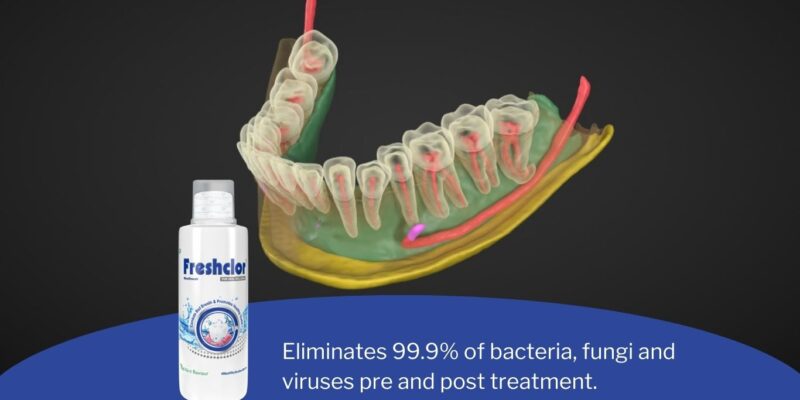
Enter AI-driven models, which promise to revolutionize this assessment by generating detailed 3D reconstructions of the mandibular canal and teeth from existing CBCT images. These models aim to provide surgeons with precise information about anatomical variations and potential risks without additional radiation exposure.
Study Overview
A within-patient controlled trial was conducted involving 25 patients who underwent bilateral mandibular third molar surgery and suffered unilateral IAN injury postoperatively. This setup allowed for a direct comparison between affected and unaffected sides within each patient, offering a unique perspective on the predictive accuracy of different imaging modalities.
Using preoperative PANO and CBCT images, researchers employed the Virtual Patient Creator AI platform to create 3D-AI models focusing on each patient’s specific anatomy. Five blinded examiners then assessed these images alongside traditional scans to determine IAN injury risk levels.
Findings
The study found that both 3D-AI models and CBCT scans had high sensitivity in predicting IAN injury risk, with scores of 0.87 for 3D-AI and 0.89 for CBCT, compared to 0.73 for PANO. However, when considering specificity and area under receiver operating curve (AUC) values—measures of diagnostic accuracy—the differences among these modalities were not statistically significant.
These results suggest that while AI-driven models perform comparably well in assessing IAN injury risk as traditional CBCT scanning—and better than PANO—they do not significantly outperform existing techniques in terms of diagnostic parameters measured in this study.
Clinical Significance
This study marks an important step forward in integrating AI into dental surgery planning by demonstrating that AI-powered 3D models can effectively evaluate IAN injury risks associated with wisdom tooth removal. The implications are significant: such technology could streamline preoperative assessments, reduce reliance on high-radiation scans like CBCT when unnecessary, and ultimately enhance patient care by providing accurate risk evaluations with existing imaging data.
Moreover, as AI technologies continue evolving, their integration into clinical practice is expected to deepen, potentially offering even greater precision and reliability in surgical planning across various medical fields.
Conclusion
The exploration into using 3D AI-driven models for assessing inferior alveolar nerve injury risks represents an exciting intersection between artificial intelligence and oral surgery. Although this within-patient controlled trial revealed no statistical superiority over traditional methods like panoramic radiography or cone-beam computed tomography in terms of sensitivity, specificity, or AUC values, it underscores the potential benefits of incorporating advanced technologies into clinical settings.
As we stand on the cusp of technological innovation within healthcare, studies like these pave the way for more sophisticated tools that promise not only improved diagnostic capabilities but also enhanced patient outcomes through personalized care plans based on precise anatomical insights.











Comments