Dr. Niharika Jain Gupta (Jabalpur, India) is a micro-endodontist and an Associate Professor at Triveni Dental College & Hospital, Bilaspur, India. She is also a research collaborate to Ph.D research projects at IIITM, Jabalpur and a key opinion leader for Micromega, Meta-Biomed, Bombay Dental & Surgical. She is one of the coveted speakers at the DRDCA 2020 International.
Introduction
Oro-facial trauma, the second most common cause of tooth loss, has a significant effect on patient’s appearance and mastication. It mainly affects children and adolescents, especially involving the crowns of maxillary central incisors. A thorough understanding of the expectation of the patient is absolutely critical for success. The repair of tooth fracture with the help of full crown or indirect veneer requires high financial expenses, is more time consuming and needs multiple appointment therapy. In this case report, single visit esthetic rehabilitation of fractured anterior tooth restored with direct veneer, after one visit endodontic therapy is presented.
Case Report
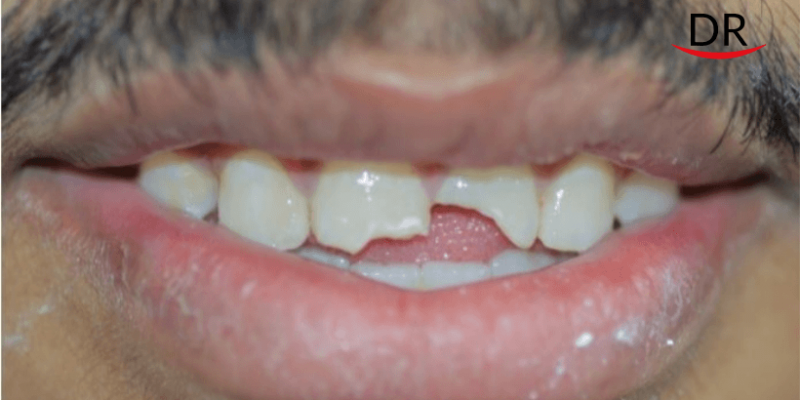
A 22-year-old male patient presented to the dental clinic, with a chief complaint of fractured maxillary central incisor caused by an accidental fall 1 month back. Thorough intraoral examination revealed Ellis Class III fracture irt 21 and Ellis class II irt 11 (Figure 1). Root canal treatment of 21 was considered as the pulp was involved. As the patient requested urgency in the treatment, a one-visit technique was planned including single visit endodontics in 21 followed by prefabricated composite resin veneer in 11 and 21.

Endodontic Procedure
Direct access was gained to the root apices under rubber-dam after local anesthetic administration. Working length was determined and glide path was established up to a size 20 K-file. Root canal preparation was done using a Self Adjusting File (four minutes) and the Vatea irrigation pump to deliver 3% sodium hypochlorite during canal preparation (Figure 2). Obturation was completed with gutta percha (F3 ProTaper Gutta Percha Cone, Dentsply Maillefer, Ballaigues, Switzerland) and calcium hydroxide based sealer (Sealapex, Kerr) using an obturating system (EQV Obturating System, Meta-Biomed, Japan)

Restorative Approach
The post space was prepared (Figure 3) to receive the fiber reinforced post. The fiber post was checked for the fit and cemented using dual cure cement [Maxcem Elite (MAX) (Kerr, Orange, CA, USA)]. Core build up was done with composite resin (Tetric N ceram, Vivadent, Lichtenstein).

Minimum Tooth preparation was done i.r.t. 11 (0.5 mm) with a diamond bur in the buccal surface to facilitate the insertion of prefabricated composite resin veneers (PCRV) (Figure 4). The PCRVs for the patient presented in this case report (Edelweiss Veneers, Edelweiss Dentistry, Wolfurt, Austria) are fabricated with a nanohybrid composite cured with high temperature and high pressure, producing a veneer with enhanced mechanical properties compared to a conventional composite restoration. In addition, these veneers are fabricated by a proprietary laser sintering technology which extracts the resin component from the composite shell and produces a veneer with high inorganic content and a highly esthetic, laser-sintered surface.

Edelweiss Prefabricated Veneers are available in four sizes (XS, S, M, L) and a custom sizing guide is provided to select the veneer which best fits the tooth preparation. For the patient in this case, medium size veneer was selected.
A diamond separating disc was used to trim the veneer and produce a sectional veneer with exact fitting. This sectional veneer was tried in its eventual position to confirm its size and shape and a retraction cord was packed around the tooth (Ultrapack size 00, Ultradent, South Jordan, UT, USA) to prevent contamination from the gingival crevicular fluid and to achieve a gentle displacement of the soft tissues. Internal conditioning of the PCR veneer was done with a resin primer (Veneer Bond, Edelweiss Dentistry, Wolfurt, Austria) as suggested by the manufacturer. This surface treatment of the veneer promotes chemical adhesion and increase bond strength between the highly inorganic composite laminate and the luting composite.
The tooth was etched with 35% orthophosphoric acid followed by the application of a single-step adhesive (G Bond, GC America) according to the manufacturer instructions. The sectional veneer was loaded with the selected composite shade, seated in position and gently pressed until it made contact with the adjacent tooth. Finally, the veneer was light cured for 20 s from the lingual direction plus 20 s from the buccal direction using a high-power curing light (Bluephase Style Light Unit, Vivadent, Schaan, Lichtenstein). Once curing was completed, the margins of the sectional veneer were finished and polished with conventional composite instruments. In the case presented in this report, a 50-micron needle point diamond bur was used to smooth the extra composite, followed by Astropol Polishing System (Vivadent, Lichtenstein) and a paper finishing strip (Sof-Lex, ESPE 3M) to polish the interproximal area (Figure 5)

Medications
Patient was instructed to take Ibuprofen 600 mg twice daily for three days and to report immediately in case of severe pain or discomfort. Oral hygiene instructions for the interproximal spaces were reviewed and the patient was rescheduled for a regular 6-month follow-up.
Discussion
Anterior tooth fracture is a tragic experience for any individual which can ultimately reduce the confidence and performance of a person in their daily activities. The search for natural esthetics and the evolution of new adhesive techniques have assured the opportunity to obtain long-term functional and esthetic results in such cases.
The design of the tooth preparation for fractured teeth can be extensive (e.g. total crown) or minimally invasive (e.g. veneers). Although different, both crown and veneer treatments require multiple clinical and laboratory steps. Contrary to this, PCRV’s have attracted a lot of attention in the dental community owing to their customization and cementation in a single session.
The prefabricated composite veneering technique has many advantages such as it is minimally invasive, preservation of natural tooth structure, chair-side technique, single appointment minimal time required to finish the treatment. The direct veneer (Edelweiss, Wolfurt, Austria) was developed in 2009 from homogeneous nano-hybrid composite using modern laser technology. These veneers are capable of mimicking human enamel and their superior mechanical properties are expanding their clinical applications.

Conclusions
Restoration of fractured anterior teeth is a complex and time consuming procedure. Excellent esthetic and functional results can be achieved with the use of a fiber–reinforced root canal post and prefabricated composite veneers like Edelweiss Direct Veneer for the treatment of anterior traumatized teeth. This advancement can be regarded as a sound investment in modern dentistry, helping a more significant number of patients to receive esthetic restorations that are more conservative and affordable.


Comments