A dental impression is a negative watermark of hard tissues teeth) and soft tissues (gingiva & mucosa) in the mouth from which a positive reproduction (cast or model) can be formed. It is made by placing an appropriate dental impression material in a stock or custom dental impression tray which is designed to roughly fit over the dental arches. Impression material is in liquid or semi-solid state when first mixed and placed in the mouth. It then eventually sets to become an elastic solid, usually takes a few minutes depending upon the material, leaving a replica of person’s dentition and adjacent structures of oral cavity.


Digital dentistry discusses the use of various technologies that incorporates digital or computer-based modules which carry out dental procedures without using mechanical or electrical tools. Digital dentistry can be used effectively for both restorative as well as diagnostic purposes. It is nowadays used as a way to facilitate dental treatments and suggest new ways to meet rising patient demands.

‘Godfather’ of Digital Dentistry is the French professor François Duret, who devised dental CAD/CAM in 1973.
TYPES OF DIGITAL IMPRESSIONS
The arena of digital impressions is quickly growing, as proved by the number of new scanner systems with more features and advantages.
There are chiefly two types of scanners on the market today.
- There are systems which use blue LED (light emitting diode). These systems are optical scanners which rest upon a reflective surface and require a contrasting medium or powder to attain a representation of the tooth morphology.
- There are systems that use laser technology to scan and measure distances from the tooth surface to secure the image. They do not entail powder.
There are four key systems on the market today :
i) Cadent iTero,
ii) 3M ESPE Lava COS,
iii) CEREC by Sirona
iv) E4D by D4D Technologies.
The Cadent and 3M ESPE machines yield models that are directed to the laboratory to produce the restoration of choice. Sirona and D4D include milling units for prompt fabrication of the final restoration chair side.
The CEREC AC can be used for fabrication of a model-only system by purchasing it without the milling centre. It utilises a blue LED digital scanner and requires the use of powder.
E4D’s system includes the interoffice milling centre to produce same-day single crowns using Culp’s database of dental anatomy as well. The difference is that E4D uses a laser scanner and it does not require powder.
ADVANTAGES AND DISADVANTAGES
Digital advantages: With milled models, we can have a solid model. Dies are precision-fit in the cast without movement, and all dies are bagged in one model. This gives greater precision when restoring multiple units, such as veneer cases. There is no contamination from the patient and no die spacer is required.
Conventional advantages: The technique has been used for many years, so it allows better initial understanding and comfort with the system is also much higher.
Digital disadvantages: Till date, there are only two implant systems on the market that allow scanning of fixture level implants — 3I and Straumann. Zimmer and Astra are the companies which believe they will have scannable abutments on the market soon. It is still difficult to scan a prepared tooth for a crown to fit an existing partial no matter which system is used.
Conventional disadvantages: Expansion and contraction of Polyvinylsiloxane material and poured casts. The possibility of bubbles, pulls, tears, and distortion while making impressions can be routinely found. There is also the fragility of stone models that require repours with less accuracy.

OTHER BENEFITS OF DIGITAL IMPRESSIONS
Digital impressions significantly increase efficacy, productivity and accuracy, and make it possible for dentists to e-mail the virtual impression to the laboratory, rather than send an old-fashioned impression or stone model through regular mail. Also, digital impressions can be used to make same day dentistry restorations, thus speeding up patient treatment and dipping the need for numerous office visits.
Other benefits of digital impressions include:
- Improved image/impression quality for better-fitting restorations.
- Reduced chair time.
- No need for unpleasant impression materials that cause some patients to gag.
- More comfortable, less apprehensive experience for patients and the dental team.
- Abridged possibility of impression-taking errors and elimination of material inaccuracies for fewer restoration mistakes.
- Patients tend get enamoured by the new technology and state-of-the-art dental care, so they become more engaged in, and better informed about, the treatment process because they can view their impressions on-screen chair side.
- The scan of the teeth being restored, as well as the opposing teeth and bite, can be done in mere three to five minutes.
- The digital impression can be stored electronically for any amount of time, which saves space, contributes to effective recordkeeping, and supports a paper-free environment.
- Green dentistry which includes jettisoning the need for disposable plastic trays and impression materials, which would be polluting landfill space and digital data is easily eliminated using the “delete” button.
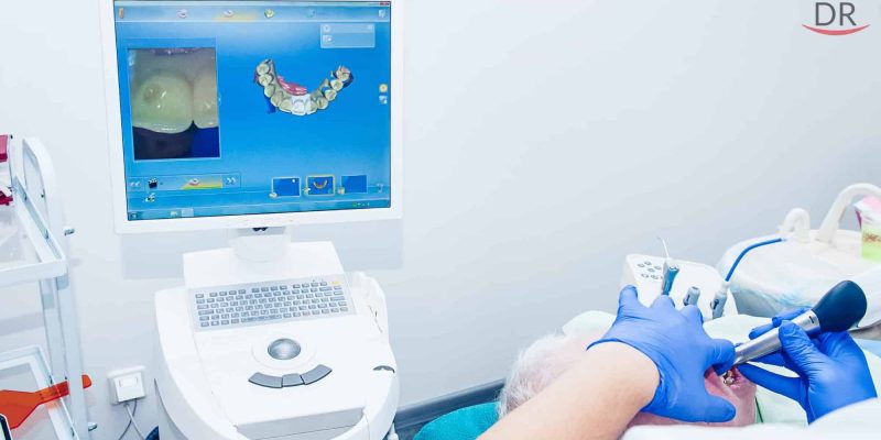
PROCEDURE OF MAKING A DIGITAL IMPRESSION
With a digital impression system, the dentist captures an image of a tooth/teeth preparation. With some optical impression systems, the area to be treated is anesthetized and made free of saliva and blood. Then the teeth are lightly dusted with specially formulated titanium dioxide powder in order to scan both arches and the bite is registered. Other systems like Cadent iTero and the impression technology that convoys the E4D Dentist System for in-office CAD/CAM restorations allow dentists to create a powder-free, three-dimensional appearance of the patient’s teeth.
The digital impression is captured by an intraoral wand which is implanted into the patient’s mouth and moved over the body of the tooth or teeth. Most of the digital impression systems use a chairside monitor which displays the impression image when it is captured.

Most of the digital impression systems also depend on point-and-click capture, which requires the images to be put together and then creating the final digital impression. However, the Lava C.O.S system is the only one which uses three-dimensional real-time video capture and then it displays live images on a touch-screen monitor. This live video capture generates the final digital impressions and doesn’t need to piece together data.
It takes approximately around a minute in capturing a digital impression of prepared teeth. An impression of the teeth in the opposite arch takes 45 seconds only.
The dentist can review the electronic real-time image, by magnifying and manipulating it for enhanced details. He can thereby ensure that whether there are any possible blunders and they can be easily corrected onscreen before sending the digital impression electronically to the dental laboratory. The delivery workflow changes depending on the digital impression system of the dental practice which it works with. Current digital impression systems available for the dental office are either digital impressions only or digital chairside CAD/CAM systems.
Digital impression systems like Cadent iTero, 3M ESPE Lava Chairside Oral Scanner (C.O.S.) and Sirona CEREC Connect are optically used to record the tooth preparations and adjacent teeth in the arch or quadrant, as well as the opposing teeth and bite registration. The data is transmitted electronically to the dental laboratory for use in restoration fabrication.
Digital chairside systems such as D4D Technologies E4D Dentist System and Cirona CEREC Acquisition Centre (AC) also record digital impressions, but the recorded data is used within the dental office in designing, milling and delivering the final restoration in one appointment for same day dentistry.
The iTero and Lava COS are the only two digital systems that can be used for all types of dental restorations, which includes bridges, crowns, inlays and onlays, and veneers. The other digital impression systems are planned so that they can be used with selected types of ceramic and composite systems.
COST OF DIGITAL IMPRESSIONS
After the initial investment which is approximately around $21,000 to $24,000(15 L to 17 L INR) for gear and training the digital scanning devices provide dental practices with abundant cost savings. They drastically reduce the requirement for conventional impression materials, which cost almost $30(2200 INR) per impression. Since files are sent electronically, there are no mailing costs; the higher accuracy and ease of digital scanning mean there are very few or almost no necessary adjustments or remakes thus representing added savings in time, efficacy, and material and labour costs.
In cases where the restoration is not considered cosmetic in nature, the cost of digital impressioning may be covered under some dental insurance plans or out-of-pocket deduction programs. Check with your insurer and dental office for specifics on your particular case.
CONCLUSION
As digital dentistry lasts to adapt and become more common, the approach to incorporate the theme of digital dentistry in learning outcomes during dental training must also change. As we enter ‘the digital age of dental education’, future practitioners requires to be exposed to new digital procedures in the curriculum and teaching so that they gain more knowledge about this newer technique.
REFERENCES
- Ting-Shu S, Jian S. Intraoral Digital Impression Technique: A Review. J Prosthodont. 2015; 24(4):313–321. doi: 10.1111/jopr.12218.
- Zimmermann M, Mehl A, Mörmann WH, Reich S. Intraoral scanning systems – a current overview. Int J Comput Dent. 2015; 18(2):101–129.


Comments