Median Mandibular Flexure
Median mandibular flexure (MMF) is a functional elastic deformation characterized by medial convergence of each half of the mandible during jaw opening and protrusive movements. The muscles, ligaments & tendons attached to mandible exert force on the bone which causes change in shape of mandible at different levels of movement of jaws.
The muscles responsible for this event are-
- Lateral Pterygoid: main muscle responsible
- Mylohyoid
- Platysma
- Superior constrictor of pharynx
These muscles, originate medially, and as a result pull the mandible medially during different kinds of jaw movement.
THE CONCEPT OF MEDIAN MANDIBULAR FLEXURE
Dental arch width and facial form are important factors for determining success and stability of orthodontic treatment. Arch form is the position and relationship of teeth to each other in all three dimensions. The mandible has a property to flex inwards around the mandibular symphysis with change in shape and decrease in mandibular arch width during opening and protrusion of the mandible. The mandibular deformation may range from a few micrometre to more than 1 mm. Median mandibular flexure mechanism (MMF) has been explained by different etiological mechanism – The contraction of the two pterygoid muscles: medial and lateral pterygoid. The distortion of the mandible occurs early in the opening cycle, and the maximum changes may occur with as little as 28% opening, or about 12 mm of mandibular movement. Median mandibular flexure is a multifactorial phenomenon. The mandible changes its shape through the extended pressure caused by the contraction of various muscles and ligament attachments.
Four patterns of jaw deformation during mandibular flexure were postulated:
- Symphyseal bending: This type of bending is associated with medial convergence or corporal approximation: this type of strain is associated with contraction of the lateral pterygoid muscle during jaw opening movements.
- Dorsoventral shear: This produces a shear force in the sagittal plane. It is a result of the vertical components of muscle forces from the lateral pterygoid muscles and the reaction forces at the condyles.
- Corporal rotation: Occurs during rotation of the body of the mandible, usually during the lower stroke of mastication. The resultant force causes narrowing of the dental arch.
- Anteroposterior shear: Occurs as a result of contraction of the lateral components of the jaw-elevating muscles. It occurs late in the power stroke, and the bending moment increases from the posterior to the anterior region.
Apart from lateral pterygoid muscle, secondary assistance from the mylohyoid, platysma and superior constrictor muscles are also responsible for the medial movement of the condyle. Mandibular flexure has also been observed during clenching, occlusion or biting forces on the mandible. Highest values of mandibular deformation occur in subjects with lower symphysis height.


RELEVANCE OF Median Mandibular Flexure IN CD PATIENTS
Median mandibular flexure poses challenging problems for both conventional and implant supported complete denture prostheses. It causes increased stress in dental implant-related prosthesis and abutments, poor fit of fixed or removable prostheses, impression distortion, pain during function, fracture of screws of implants or porcelain crowns, loosening of cemented prostheses, and resorption around implant.
Considerations in impression making
The medial convergence of the mandible due to contraction of the muscles by the conventional open mouth impression technique may alter the accuracy of the master cast and result in compromised prosthesis. Moreover, when impressions are obtained with mandible at a position of maximum mouth opening, the teeth will be recorded in a more lingual position than they would actually be found at rest or in occlusion. Impressions made using closed-mouth technique result in minimal activation of the muscles of mastication and help to reduce the amount of mandibular deformation. Impressions made with mouth wide open may lead to incomplete seating of fixed prosthesis that may later require extensive occlusal correction.
Considerations while recording centric relation (C.R.).
Mandibular flexure occurring either during patient guided C.R. registrations or during function may influence the fit of the prosthesis and make it difficult for the clinician to achieve good occlusal contact. This is further complicated by the use of rigid dental casts mounted within rigid articulators. This often results in severe discrepancies as a closed-mouth C.R. record may not accurately fit onto a dental cast made from an open mouth impression technique. Secondly, the framework of a fixed dental prosthesis fabricated on a cast made from an open mouth impression technique would not fit accurately in patients’ mouth. Such prosthesis would have occlusal interferences and result in pain when the patient applies a biting force. Therefore, a ‘closed-mouth’ impression technique and a dentist-guided C.R. recording technique should be used to reduce such discrepancies.
Considerations while occlusal mounting.
Mandibular flexure in the horizontal plane often results in discrepancies between cusp tip indentations in the jaw registration records and cusp tip location on the dental casts. The articulation of the casts is affected due to the lingual movement of mandibular teeth. This type of occlusal mount will not represent the correct occlusal relationship. The restorations fabricated to such recordings could present with occlusal interferences and articulators may require modification so as to allow for mandibular resilience.(1)
Median Mandibular Flexure AND PERIODONTAL CONSIDERATIONS
The narrowing of the mandibular arch abnormally stimulates periodontal ligament mechanoreceptors with significant influence on the health of the periodontal apparatus and surrounding alveolar bone. A rigid splinting is commonly performed on periodontally involved teeth where half to two-thirds of the alveolar bone support is lost. Such a rigid fixed splint will restrict the movement of the mobile teeth. In such a situation, the teeth fail to move when the mandible moves in both linear and rotational directions during flexure. This creates stress in the periodontal attachment and causes increased tooth mobility with alveolar bone loss.
RELEVANCE OF Median Mandibular Flexure IN FPD
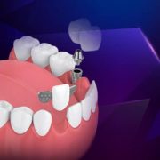
Roots from dental implants have become an accepted option for the treatment of dentulous and partially edentulous patients. For patients requiring replacement of teeth in posterior quadrants, implant-supported restorations provide alternatives to distal extension removable partial dentures and distal extension cantilever fixed partial dentures. The use of tooth-supported cantilevers should be avoided in posterior regions because of the magnitude and direction of occlusal forces that are placed on associated pontics. When only one implant successfully integrates in a posterior edentulous space or when there is insufficient space to place more than one implant, a dentist may consider restoring a single implant.
This implant can serve as an abutment for a single crown, or it can be combined with an adjacent natural tooth to provide support for a fixed partial denture. Combining an implant and a natural tooth abutment within a single fixed partial denture may be a solution to the problems associated with the restoration of a unilateral posterior edentulous area in which only a single implant can be used.(3)


RELEVANCE OF Median Mandibular Flexure IN IMPLANT
Mandibular flexure potentially affects the accuracy of different stages of implant treatment, including osseointegration of the surrounding bone, implant prosthesis fabrication, strain distribution within the framework during mastication, crestal bone around implants, etc. The flexural forces cause lateral stresses on the implant body resulting in bone loss around implants, loss of implant fixation, material fracture and discomfort on mouth opening.
Therefore, it is essential to consider MMF while planning any implant-supported prosthesis. Median mandibular flexure causes microdamage at the crestal region and poor osseointegration due to micromovements around implants. Fischman (1990) explained the importance of rotational aspect of mandible on the osseointegration of implants. Hobkirk and Schwab (1991) have also confirmed that the posterior implants could be subjected to stress-induced microdamage at the bone implant interface in cantilever situations due to mandibular flexure. A relative displacement of up to 420 microns and force transmission of up to 16 N between linker implants with jaw movements is possible during mandibular flexure. The forces were more during opening and protrusive movements than lateral excursion.9
A linear distance of implants which had been placed 10 mm apart showed more variation in distance during protrusion compared with maximum mouth opening position. Hobkirk and Havthoulas (1998) studied the influence of functional mandibular deformation on the force distribution in the jaws, implant or superstructure complex. The results showed that the force distribution in the mandibular implant host complex is unevenly distributed about the median sagittal plane as a result of jaw asymmetry and an increase in the number of implants that support a fixed superstructure would results in a leverage effect around the midline.
WHAT HAPPENS IF MEDIAN MANDIBULAR FLEXURE IS LOST?
Certain situations like trauma or surgical resection due to benign and malignant neoplasms may cause disruption in the continuity of the mandible. Usually the most common type of resection is hemimandibulectomy where one half of the mandible is resected, with or without muscles. Loss of mandibular continuity causes a loss of median mandibular flexure. In normal patients, during opening – there is a decrease in mandibular arch width and protrusion of the mandible because of flexure. In hemimandibulectomy patients, during opening – there is deviation of the remaining mandibular segments towards the defect because of loss of flexure and rotation of the mandibular occlusal plane inferiorly because of scar contracture and an imbalance in the muscular system causing a more inferior muscular pull . This irregular movement of mandible i.e. deviation and inferior rotation has a profound influence on normal form and function. It results in facial disfigurement, loss of occlusal contact, slurry speech, improper mastication, in many cases, loss of lip competency for saliva control and to initiate the swallowing process.
CONCLUSION: Median Mandibular Flexure
Mandibular flexure is a multifactorial phenomenon that occurs instantaneously and concurrently with jaw movements. The mandibular flexure can affect the prognosis and treatment outcome for various dental and implant-related procedures. Therefore, it is very important to take appropriate measures and adopt correct technique that helps negate the flexural movement of the jaw during any prosthetic rehabilitation. This would not only help clinician to achieve good prosthesis with accurate fit but also help to maintain the health of the surrounding periodontal and osseous tissues.
REFERENCES
- Osborne J, Tomlin HR. Medial convergence of the mandible.Br Dent J. 1964;117:112–114.
- Novak CA. Mandibular dimensional change in various jaw positions and its effect upon prosthetic appliances. DentStud. 1972;50:19.
- Sivaraman K, Chopra A, Venkatesh S. B. Clinical importance of median mandibular flexure in oral rehabilitation: a review. Journal of Oral Rehabilitation.2016; 43:215-225.
- Lingeshwar, Appadurai. Prosthodontic management of hemimandibulectomy patients to restore form and function – A case series. World J Clin Cases. 2017 Oct 16; 5(10): 384–389.


Comments