Dr Nikhil Bahuguna was the Mentor of the DR Pronto Esthetic Challenge 1, 2021-2022


Introduction
Dental trauma in young adults is one of the most common reasons for seeking emergency treatment.

Injuries can range from a simple enamel infarction, to fracture of teeth exposing the pulp-dentin complex, to avulsion of the tooth.
The treatment choices depend upon the amount of tooth involved in trauma and in case of fracture of a fragment or avulsion, the time lapse from the accident to the replantation or the reattachment of the fractured or dislodged tooth. Additionally, of considerable importance, is the storage medium of the dislodged fragment or tooth.
Case report
A young boy, 13 years reported with complaint of trauma to the anterior upper teeth region.
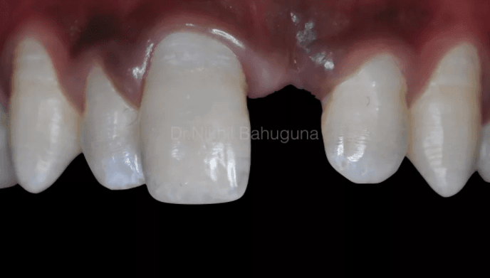
On examination, the upper left central incisor (21) was avulsed and lost; and right central incisor (11) had an Ellis class 4 fracture – involving the pulp.


The time lapse between trauma and reporting for the treatment was about 2 hours, so it was considered as a favorable time for the proposed treatment.
The treatment was carried out in the following steps –
1. Socket cleaning with betadine and slough cleaning, followed overall disinfection of the trauma site.
2. IOPA was done to evaluate the status of 11. No obvious root fracture was as observed
3. Rubber dam isolation was achieved.

4. 11- the exposed pulp tissue was removed from the coronal chamber, pulpotomy was performed retaining the radicular pulp.

5. The pulp tissue was cleaned with normal saline and observed for cessation of any frank bleeding.
6. Mineral trioxide aggregate (MTA+, Cerkamed) was used to seal the exposed radicular pulp, covered with a moist cotton pellet to aid in setting and covered with a pre mixed temporary cement. Patient was prescribed necessary medication and recalled after 24 hours.


IOPA was taken to verify MTA placement at orifice level.

7. After 24 hours, rubber dam isolation was achieved and the temporary restoration was removed and MTA set verified.

8. The tooth was then prepared. Buccal and palatal surface beveling was done for surface extension.

9. The fractured fragment (stored overnight in cooled saline) was then prepared.


10. The pulp chamber was cleaned, tooth treated with 5% NaoCL, buccal and palatal bevels and chamber slots were prepared followed by etching and bonding.


The buccal and palatal surface beveling of the prepared tooth surface was etched and bonded intra-orally.

11. Mineral enriched light cure stackable composite, Active™ Pronto (A2) was used as a bonding medium to attach the two fractured fragments together.

12. Activa™ Pronto was also used to cover the buccal and the palatal bevel extensions for good adaptation and a better surface seal.

13. The surface was covered with an oxygen inhibition gel and cured again.

Notice the immediate post-op view showing a clean reattachment of the fractured fragment.


14. Initial finishing was performed with red grit diamond points, and cups and cones (Enhance) after 15 minutes.

15. Final polishing was done after 48 hours with cups and cones (PoGo) and diamond polishing paste and buffs.

16. A fibre bonded bridge was then done with a direct build up using multi directional pre adhered glass fibers for pontic fabrication and reinforced with a glass mesh for better strength (UFM Fibre, Dentapreg). The pontic was fabricated using enamel and dentin effect shades (Neo Spectra range). Find the steps below:


17. Final finishing and polishing was then done and case recalled after 6 months.


18. 7 month recall showed stable results. IOPA showed no significant peri-apical or periodontal changes.


The current work flow exhibits, one of the many options for management of fractured teeth where the fragments can be re attached using flowable / stackable mineral enriched composites like Activa™ Pronto and fabricating a direct bonded bridge using fibers and histology based composite resins.
You can find out more information about flowable / stackable mineral enriched composites here.


Comments