Introduction
Studies have shown that 1 out of every 4 persons under the age of 18 will sustain a traumatic anterior crown fracture.1,2 Such fractures especially in the esthetic zone create serious functional, cosmetic and psychological problems for both children and their parents.
The main challenge for a clinician while managing such patients is to choose a treatment that will return the tooth to its original form. There are various modalities of management of fractured anterior teeth:
- restorative build-up of the fractured segment,
- post & core
- crowns
- fragment reattachment
Reattachment of fractured tooth fragment when its available, is one of the best options for managing a fractured anterior tooth. It offers a quick, economic and conservative solution that restores the form and function of the tooth in the same appointment.The successful reattachment of the fragment primarily depends on the retrieval of the fragment at the time of injury.
Case Report
A 9-year-old girl was brought to our clinic by a very disturbed mother explaining in detail, how her child had suffered trauma due to a fall while playing. She informed that the incident occurred 10 minutes ago and she brought the child to the clinic immediately. At this point, seeing her fractured upper anterior teeth, we requested the mother to go back to the site of the incident and look for any broken pieces of tooth and bring them in a cup of milk as soon as she can. Meanwhile, we took care of her daughter’s injuries and made her comfortable at the clinic.

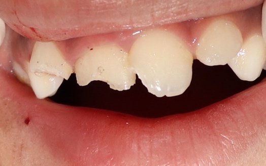
Figure 1: Pre-Operative
The child was responsive, presenting a calm and collected demeanor. She was oriented in time, place and person after injury. Detailed medical and dental history was recorded.
On extra oral examination, visible bruising could be seen over her lips, cheeks and chin. There was superficial bruising over her legs, knees and elbows.
Intra Oral examination revealed Ellis Class II fracture of 12, 11 and 21. The teeth were tender on vertical percussion with grade 1 mobility.
All her external injuries were cleaned with saline and disinfectant.
In the meanwhile, her mother returned with the pieces of tooth salvaged from the site of the incident. Among the pieces, only one big piece could be used with 11 which was intact. The rest of the pieces were too small to be approximated with the rest of her teeth.

Figure 2 : Fragment Approximation

Figure 3 : Fragment Fit
A pre-op IOPA revealed involvement of only enamel and dentin with 12, 11 and 21.

Figure 4 : Pre-Op IOPA
It was decided to re-attach the fragment to 11 and build up 12 and 21 with composite resin. The patient’s family was informed of the same and her mother was very happy to have her child’s original tooth re-attached.
Fragment re-attachment
The fractured tooth segment was positioned back to the normal position for 11 apposing their fracture lines and luted with flowable composite (Activa Pronto, shade A2) after etching and bonding (Prime and Bond Universal Adhesive System, Dentsply).

Figure 5 : Priming of the fragment
No bevelling was done to the original tooth. The fractured fragment was treated with adhesive (Prime and Bond Universal Adhesive System, Dentsply) and simply bonded.

Figure 6 : Etching with 37% Phosphoric Acid

Figure 7 : Curing of Adhesive
12 and 21 were restored with restorative resin (Neospectra packable composite A2).
Finishing and polishing was done (Shofu Super Snap Discs and 3M Spirals).

Figure 8 : Post Operative

Figure 9 : Immediate Post-Op IOPA

Figure 8 : Palatal View

Figure 11 : Post Op After 7 days (Turmeric application by mother caused the yellow discoloration)
During follow up visit after one week, it was observed the tooth was periodontally healthy and no fracture line was noticeable.
Follow- up clinical evaluation was conducted 6 months later, which revealed 11 to be esthetically and functionally fine.
Discussion
Advancements in the field of adhesive dentistry allow the clinicians to reconstruct the natural tooth of the patient in a minimally invasive way achieving both, esthetic and functional requirements.
The technique of fragment reattachment was pioneered by Chosak and Eidelman, when they first published in 1964, a case involving the reattachment of a natural tooth fragment.They used a cast post and conventional cement to reattach an anterior crown segment on a 12-year-old boy.3
The success of reattachment depends on certain factors like
- the site of fracture
- size of fractured fragment
- periodontal status
- pulpal involvement
- status of the root formation
- biological width invasion
- occlusion
- time passed since trauma
- fragment hydration and
- materials used for reattachment.4
The amount of time passed after trauma and the fragment remaining hydrated played an important role in the success for this case.
Milk was chosen as a storage media due to its ease of availability as it has better physiologic properties, including pH and osmolality.5 Milk was used in a study as the hydration media to test its effect on fracture resistance. After hydration of tooth fragment for time interval of 1hr, 6hrs and 24 hrs, it showed effective fracture resistance when compared with dry storage.6
Change in dental colour due to dehydration of the fractured fragment leads to decrease in fracture strength of the tooth. A well rehydrated fragment has the capability of restoring both color and strength. Farik et al., in his studies observed that additional drying of fractured fragment beyond 1 hour decreases the fracture resistance significantly, thus concluding the importance of keeping the fragment moist.
Shirani et al, concluded that 50% of the fracture strength of the original tooth is restored with hydration or without dehydrating of the surfaces with storage medium and without any additional preparation. 7
Hydration maintains the vitality and original aesthetic appearance of the tooth 8. Farik et al, have explained that etched, rinsed, but not dried dentin has a surface of partly demineralized hydroxyapatite which is covered with un-collapsed collagen fibers.
Dentin bonding agents when applied on this surfaces creates a bond by mechanical interlocking with collagen and fill the space in the partly demineralized hydroxyapatite on polymerization. If the etched and rinsed surface is dried to a certain extent, the collagen fibers will collapse and prevent the bonding agent penetrating to the partly demineralized zone.
This results in relatively low bond strength and therefore may have important consequences for the reattachment procedure for fragments.9

Figure 14: Post Op IOPA after 6 months showing apical closure
Advantages of Re-attaching the fractured fragment:
1. Wears at the same rate as that of natural tooth

Figure 12 : Post Op 6 months Lateral View
2. Enhanced esthetics due to natural enamel translucency and surface finish

Figure 13 : Post Op 6 months Frontal View
Hence, re-attachment stands to be an effective, economical and conservative procedure to restore the natural original shape, contour, translucency, surface texture, occlusal alignment, and colour of the fractured tooth resulting in a positive emotional and social response from a patient.10,11
Conclusion
The high incidence of trauma to anterior teeth calls for quick and esthetically functional strategies for management. The age of the patient plays a significant in handling the individual and in such cases, re-attachment procedures emerge as a fast and effective way of treatment provided the fragment is available.
The main concern is that the general public remains unaware regarding this treatment strategy and most patients report say that they discarded the fragment when the trauma occurred. Our role as dentists should be to create awareness in the community on retrieval of the fragment and to seek immediate dental care and some tips on how to bring it to the dental office.
References
1. J. O. Andreasen and J. J. Ravn, “Epidemiology of traumatic dental injuries to primary and permanent teeth in a Danish population sample,” International Journal of Oral Surgery, vol. 1, no. 5, pp. 235–239, 1972.
2. S.PettiandG.Tarsitani,“Traumaticinjuriestoanteriorteethin Italian schoolchildren: prevalence and risk factors,” Endodontics and Dental Traumatology, vol. 12, no. 6, pp. 294–297, 1996.
3. [d1] Chosack A, Eidelman E. Rehabilitation of a fractured incisor using the patient’s natural crown: Case report. J Dent Child 1964;31:19-21.
4. C.P.K.Wadhwani, ―A single visit, multidisciplinary approach to the management of traumatic tooth crown fracture,‖ BritishDental Journal, vol. 188, no. 11, pp. 593– 598, 2000.
5. [d1] Bazmi BA, Singh AK, Kar S, Mubtasum H. Storage media for avulsed tooth- a review.Int Journal of Multidisciplinary Dentistry 2013;(3):741-744.
6. Rahul HJ, Swati KJ. Comparison of the Effect of Various Storage Media on the Fracture Resistance of the Reattached Incisor Tooth Fragments: An in vitro Study. Indian Journal of Dental Sciences 2017;9(4):233-236.
7. Shirani F, Malekipour MR, Sakhaei Manesh V, Aghaei F. Hydration and dehydration periods of crown fragments prior to reattachment. Oper Dent 2012;37:501-8.
8. Capp CI, Roda MI, Tamaki R, Castanho GM, Camargo MA, de CaraAA. Reattachment of rehydrated dental fragment using two techniques. Dent Traumatol 2009;25:95-9.
9. Farik B, Munksgaard EC, Andreasen JO, Kreiborg S. Drying and rewetting anterior crown fragments prior to bonding. Endod Dent Traumatol 1999;15:113-6.
10. [d1] R. D. Trushkowsky, ―Esthetic, biologic, and restorative considerations in coronal segment reattachment for a fractured tooth: a clinical report,‖ The Journal of Prosthetic Dentistry, vol. 79, no. 2, pp. 115–119, 1998.
11. 9. F. C. S. Chu, T. M. Yim, and S. H. Y. Wei, ―Clinical considerations for reattachment of tooth fragments,‖ QuintessenceInternational, vol. 31, no. 6, pp. 385–391, 2000.
Author – Dr Urvashi Tanvar





Comments