It has been found that with progression of age-related macular degeneration is the leading cause of deteriorating eyesight in people over 50 is hastened by deposits of a mineral found in tooth enamel at the back of the eye.


Amelotin may turn out to be a therapeutic target for the blinding disease. These findings were published in the journal Translational Research. NEI is part of the National Institutes of Health.

“Using a simple cell culture model of retinal pigment epithelial cells, we were able to show that amelotin gets turned on by a certain kind of stress and causes formation of a particular kind of calcium deposit also seen in bones and teeth. When we looked in human donor eyes with dry AMD, we saw the same thing,” said Graeme Wistow PhD, chief of the NEI Section on Molecular Structure and Functional Genomics, and senior author of the study.
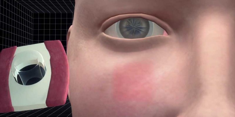
The deposits themselves are composed of mineralised calcium called hydroxyapatite which is also a key component of tooth enamel and bone. When these deposits occur under the retinal pigment epithelium, they can impair vision by blocking the access of oxygen as well as nutrients to the photosensitive cells of retina. This results in significant distortion of a person's vision. Small balls of hydroxyapatite filled with cholesterol, called spherules, were found only in people with dry AMD, and not in those with wet AMD or without AMD.

OOK Prosthesis – A Tooth for Restoration of Vision! – Read here
“Prior to this study, nobody really knew how the hydroxyapatite was accumulating in the dry AMD drusen,” said Dinusha Rajapakse PhD, the first author of the study. “Finding this tooth-specific protein in the eye, this protein that’s linked to hydroxyapatite deposition – that was really unexpected.”
During the experiments, researchers grew retinal pigment epithelial cells in the laboratory and then forced them to starve blocking access to nutrients. Nutrient deficiency lead to the production of amelotin, which, in turn contributed to the formation of calcium deposits. Blocking the production of amelotin stopped the formtaion of deposits.
“Mechanistically, amelotin looks like a key player for the formation of these very specific hydroxyapatite spherules. That’s what it does in the teeth, and here it is in the back of the eye. Conceptually, you could see coming up with drugs that specifically block the function of amelotin in eye, and this might delay the progression of the disease. But we won’t know until we try it,” said Wistow.
“It’s a ways off, but the best possible outcome would be finding a way to inhibit amelotin or its gene expression in a way that prevented or delayed the deposition of hydroxyapatite,” Wistow says. That “might allow people to keep their vision a bit longer.”
Source: ScienceDaily.com


Comments