Just remember the acronym “ORALSURGERY”!


1. “O”- Omission
Before you learn to choose your extraction cases, you need to learn which cases you should not do. Selecting cases very carefully and thoroughly as first success and failure would become foundation for your lifelong confidence and your image.

For example,
- Do not take any patient with extreme medical illness (ASA category 3 or 4). To learn more about this please click here !
- Deep mesioangular lower 3rd molar would be easier to extract than fully erupted distoangular lower 3rd molar tooth.
- In case you are not sure of the diagnosis, then do not continue with the extraction just because the patient is insisting or willing to go for it. (In most of the trigeminal neuralgia cases, patients often force the dentists to do extractions at the site of external pain, and later themselves blame the dentists for the same.)
2. “R” – Roentgenize
It is better to have an X-ray of almost all the extraction you are planning in the initial stage itself, irrespective of their eruption status or their mobility status.
- Erupted teeth with curved roots will be difficult to extract than impacted but with short conical roots.
- There might be a periapical cyst or tumor which may lead to unexpected intra-operative complications which can be easily avoided by having a knowledge of its existence through X-ray beforehand.
- For medicolegal reasons, it is better to have an investigation done before treatment and kept as record/evidence.
How to roentgenize?
- Go for IOPA if planning extraction of erupted tooth.
- Go for OPG if you are planning for extraction of an impacted lower 3rd molar extraction and
- Go for CBCT if the roots of lower 3rd molar to be extracted are in close approximation with inferior alveolar nerve canal as per IOPA or OPG.
3. “A”- Anesthesia
Here are few tips on how to ensure effective and painless anesthesia:
- Apply topical LA over the injection site so that needle insertion is not painful, especially for anxious and pediatric patients.
- Increase the dose to compensate for the skills: initially your anesthesia skills are still under refinement stage, so it is better to give multiple time injections rather than giving just once or twice. Just respect the maximum recommended doses 4mg/kg of the body weight.
- Intraligamentary injections can be added along with nerve blocks or infiltrations as it doesn’t add much to the dose and still it helps in achieving pain control.
- For patients with cardiac diseases, it is better to use plain local anesthesia (without adrenaline) or else limit the dose to 4 cc only.
- Buffered local anesthesia is a better choice in patients with previously failed anesthesia or patients with oral submucous fibrosis.
What is ‘Buffered LA’?
Buffered local anesthesia is adding alkalizing agent to the local anesthetic solution before injecting .The most common method for buffering of local anesthetics is with the addition of sodium bicarbonate – an alkalinizing agent, which is most used for the treatment of metabolic acidosis. 1.0 ml of 8.4% sodium bicarbonate is added to every 9 ml of local anesthetic solution (1:9). Directly load from the sodium bicarbonate ampule to the syringe and then load LA – or otherwise. The injection technique remains the same, its only the LA agent which is modified.
Alkalinization or buffering of dental local anesthetics to raise the pH of acidic LA solutions is a well-documented technique that results in clinical benefits such as
- decreased injection pain
- reduced onset time
- need for less overall volume of local anesthesia.
4. “L” – Larger the better
Whenever planning to give incision, it is better to give bigger incision than the smaller incision as visibility will be better and the final healing time will be always the same.
5. “S” – Stabilize
It is particularly important to stabilize the mandible while extracting lower teeth or else it may lead to temporo-mandibular joint disorders. You can stabilise with the fingers of your other hand. If bite block is used along with fingers to stabilize, it will be an added advantage!
6. “U”- Unknown
There can be many unknown things about the patients which even patient does not know and for that you need to advise either blood investigations or deep interrogations.
For example:
- Patients above 40 years should be investigated for diabetes mellitus as it directly affects healing of extraction wounds.
- All (married) females of reproductive age group should be asked for history of pregnancy, as in INDIA patients often hesitate to talk about pregnancy to the treating dental surgeon.
- All patients above 60 years should be asked about the history of chest pain or medications for the same.
- All patients should be investigated for coagulation profile if they are on oral anticoagulants, or they have had history of cardiac related surgeries.
- All mentally retarded patients (or caretakers) should be asked or investigated to rule out epilepsy.
- All patients with spontaneous bleeding in the oral cavity from the gingiva, in the absence of significant dental plaque and calculus should be investigated to rule out leukemia.
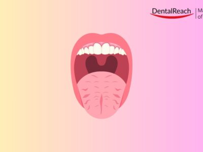
7. “R” – Rima oris
Rima oris is nothing but the oral aperture width. Patients with adequate mouth opening may have small oral aperture which may be either physiological/ anatomical and or pathological (e.g., oral submucous fibrosis / scleroderma/ burns/ post tumor resection surgical fibrosis/ radiation induced fibrosis). This would lead to the difficulties related to the retraction, accessibility and visibility intraorally. Kindly note here the interincisal mouth opening is near normal. Although there is no objective method to record and assess the normal oral aperture width, one can simply check by looking at the full smile of the patient, which should be minimum up to the level of upper or lower molars. The decreased rima oris will result in the reduced retractability of the cheeks which ultimately will result in decreased accessibility and visibility.
Following modifications can be tried to manage decreased or stiff rima oris:-
- Give larger relieving incision and raise larger flap – avoid envelope flap.
- Use thicker and broader retractor like Minnesota retractor.
- Aggressive bone removal.
- Consider giving steroids perioperatively as in such patients the chances of soft tissue trauma and edema are higher.
8. “G”- Gingerly
Go slow. It’s a myth that the faster you extract, the better you are. It is better to go slow!
9. “E”- Elevate with proper care.
The right technique of elevation can pop the tooth out of the socket in seconds! Read the DentalReach article for more details.
10. “R”- Recumbent
Recumbent position is defined as ‘lying down, especially in a position of comfort or rest; reclining’. While supine position is defined as lying on the back or having the face upward, it can be supine, right lateral and left lateral positions.
In case of loss of consciousness, immediately position the patient in recumbent position. Which recumbent position you choose, depends on certain factors:
- In general, all the patients with loss of consciousness should be positioned in supine position.
- All pregnant patients should be positioned in the left lateral position not in the supine position to prevent Supine Hypotensive Syndrome of Pregnancy.
- If the patient has swallowed foreign body, then also lateral recumbent position is preferred.

11. Bonus tip! “Y”- Yours only
Follow above mentioned 10 tips for safe and smooth oral surgery, as the responsibility of the safety of the patient is: —–
“Y” yours only! Whatever a physician or the patient says, the ultimate responsibility of the case medico-legally is yours only so do not trust physicians or the patients blindly. Take proper informed consent before the procedure and then go ahead with surgical extractions.


Comments